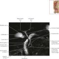
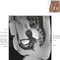
Penis and Male Urethra Axial 1
Diagnostic Consideration
Common indications for magnetic resonance imaging (MRI) of the penis include detection and staging of penile and urethral malignancies, evaluation of penile prostheses, and detection of penile fractures, Peyronie’s disease, and periurethral abscesses.


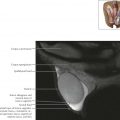
Penis and Male Urethra Axial 4

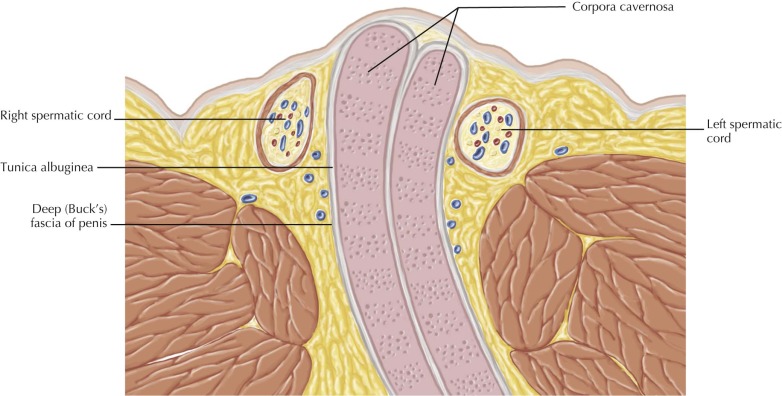
Normal anatomy
The tunica albuginea is a thin layer of fibrous tissue that surrounds each of the corpora cavernosa and the corpus spongiosum. The deep (Buck’s) fascia is just superficial to the tunica albuginea and surrounds the corpora cavernosa in a dorsal compartment and the corpus spongiosum in a separate, ventral compartment. The tunica albuginea and deep (Buck’s) fascia are inseparable on MRI and appear as a single, hypointense border surrounding the corpora. The superficial (dartos) fascia is seen more superficially, just beneath the skin and subcutaneous fat.


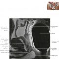
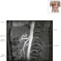
Penis and Male Urethra Coronal 1