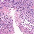
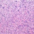
Histologically the tumor is very similar to enchondroma, but it more frequently displays features of cell proliferation (high cellularity, nuclear plumpness, and frequent double-nucleated cells). Being somewhat painful and causing some swelling in most instances, it usually requires surgical management consisting of either en bloc marginal excision or thorough curettage, equally effective.
Key Points
Clinical | Some pain, young patients |
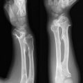
Radiological | Subperiosteal, metaphyseal, with erosion of the cortex, granular calcifications |
Histological | Lobules of benign cartilage. Possible hypercellularity |
Differential diagnosis | Periosteal chondrosarcoma |
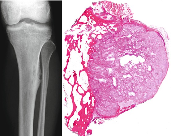
Male, 22 years old. Small (less than 3 cm) well-circumscribed lobulated lesion composed of hyaline cartilage. The lesion is beneath the periosteum with a sharp margin with the underlying cortex. The chondrocytes frequently are enlarged and hyperchromatic with increased cellularity and variability in nuclear size and shape. Out of context, these features suggest the diagnosis of chondrosarcoma
Related posts:


Stay updated, free articles. Join our Telegram channel

Full access? Get Clinical Tree


