Imaging: On x-ray – typical aspect of an osteochondroma with more abundant calcifications or ossifications, intense, and diffused radiopacities, with thicker superficial uncalcified layer and with fuzzy margins towards the soft tissue. At times, the implant base of the osteochondroma is still visible or the tumor invades the medullary canal. On bone scan – intense uptake. On CT – many, large, noncalcified tumoral lobules, ringlike, popcorn-like radiopacities, and higher thickness of the cap of cartilaginous tissue. On MRI – lobulated, ill-defined, inhomogeneous muscular signal intensity on T1, extremely heterogeneous signal intensity on T2 with large foci of signal void due to the calcified areas, and with a thick peripheral layer of white signal due to the cap of the lesion.
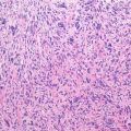
Histopathology: Large, bumpy, cauliflower-like, with a thin pseudocapsule and thick cap of cartilage. Cartilage involves the spongy bone of osteochondroma; it is softer, juicy, grayish, and translucent. Calcifications are very intense in low grade C.: granules, rings, spots of yellowish-white aspect, chalky and gritty, with hard consistency. Lobules of well-differentiated cartilage: many large cells in groups with abundant matrix, with double hyperchromatic nuclei. Formation of nodules, mitosis, and cystic to myxoid changes may be seen. Nests of tumoral cells are observed in the pseudocapsule. Cartilage infiltrates the marrow spaces of cancellous bone of the osteochondroma.
Course and Staging: It grows more slowly than central C, recurrence is observed from a few months to 10 years, <20 % of cases have lung and late metastases. Dedifferentiated peripheral C is rare (4 %). Usually, stage IB.
Treatment: Wide resection. Inadequate surgery may cause scattered neoplastic nodules in scarring tissue. Radio- and chemotherapy are not effective. Amputation is necessary when it is too large and otherwise inoperable. Peripheral C is less malignant than central because grade 1 forms are frequent and grade 3 rare.
Key Points
Clinical | Slow increasing swelling
Related posts:Stay updated, free articles. Join our Telegram channel
Full access? Get Clinical Tree
 Get Clinical Tree app for offline access

|