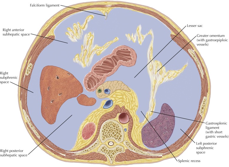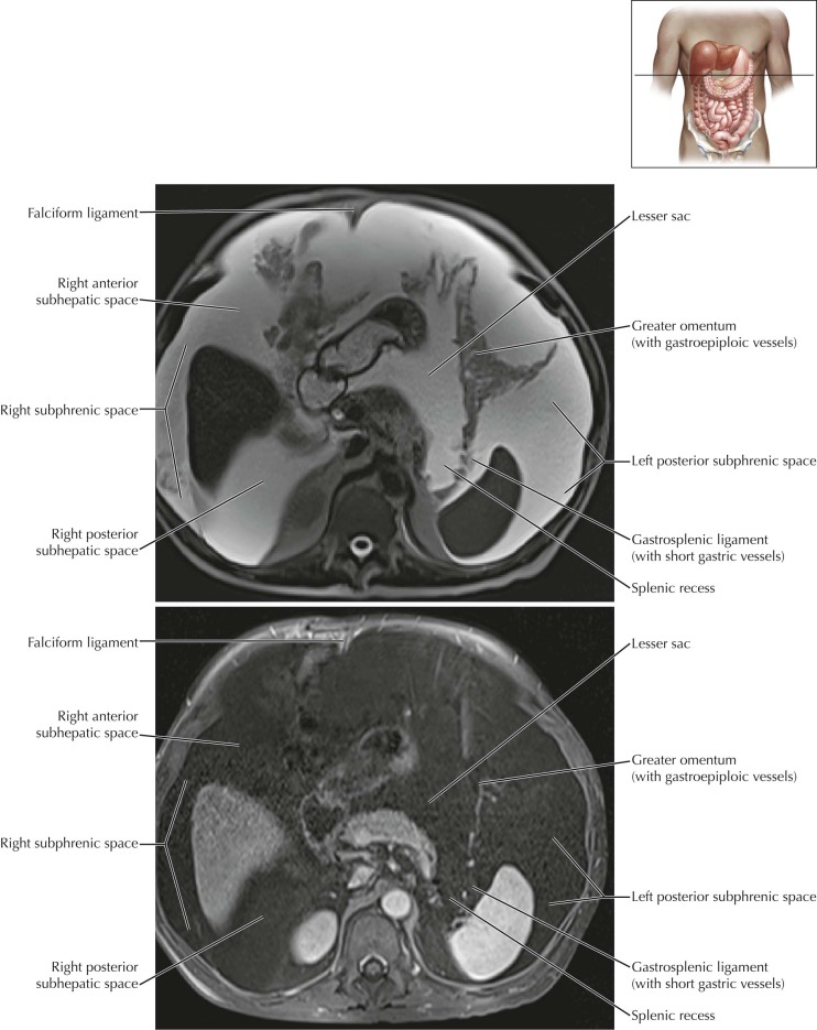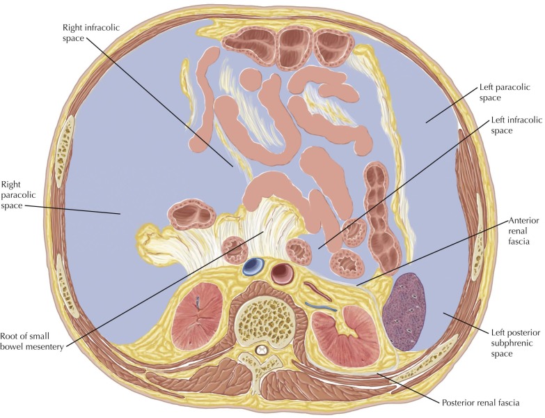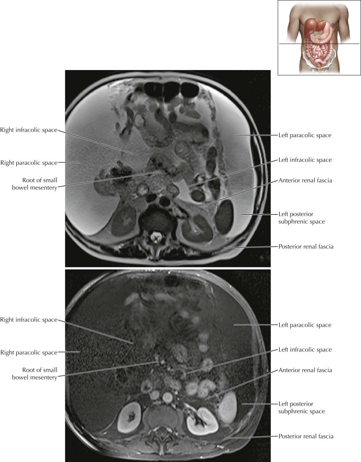
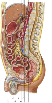
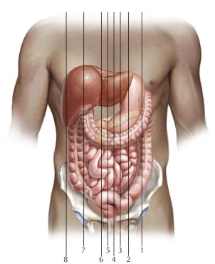
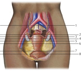
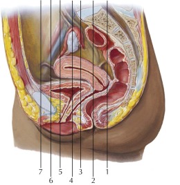
Peritoneal Cavity–Abdomen Axial 1
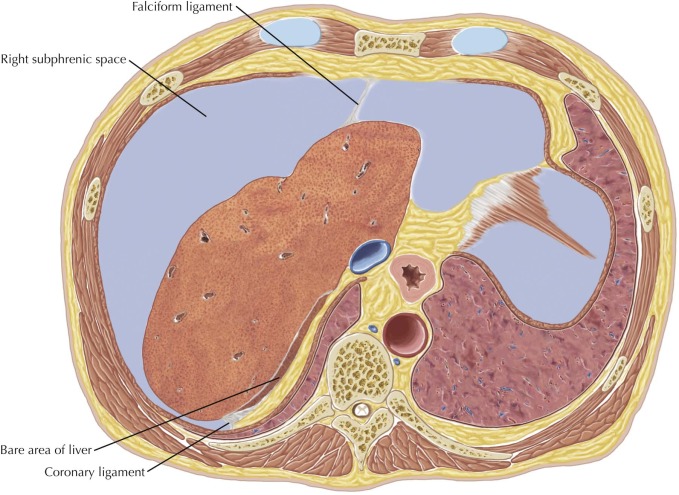
Normal anatomy
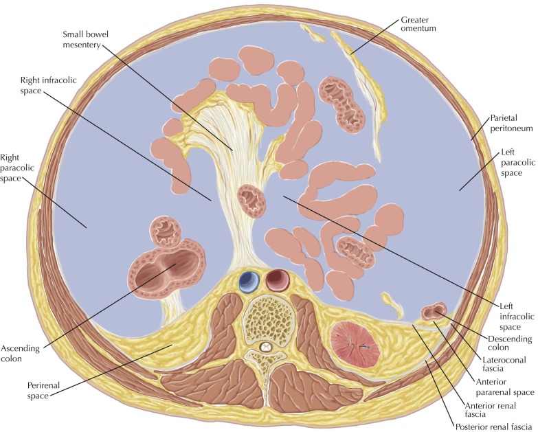
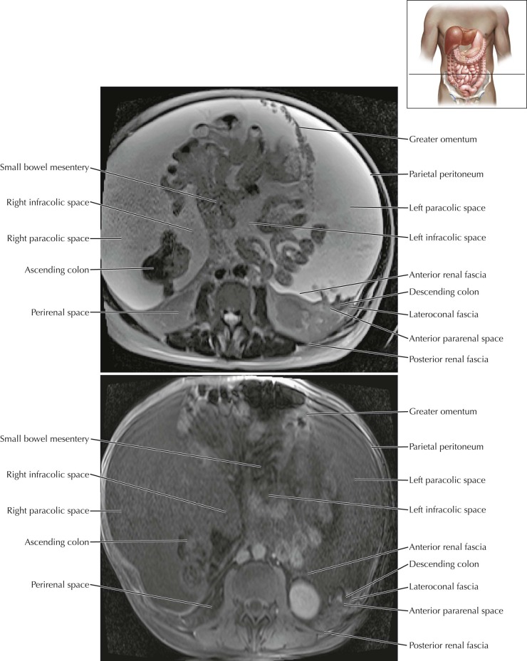
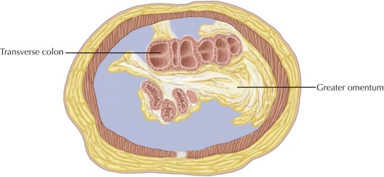
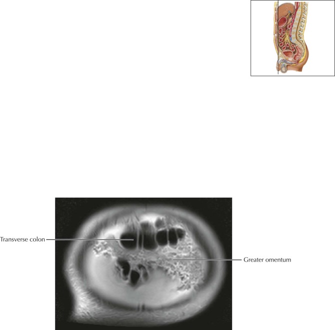
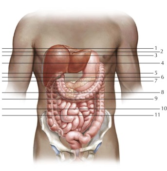
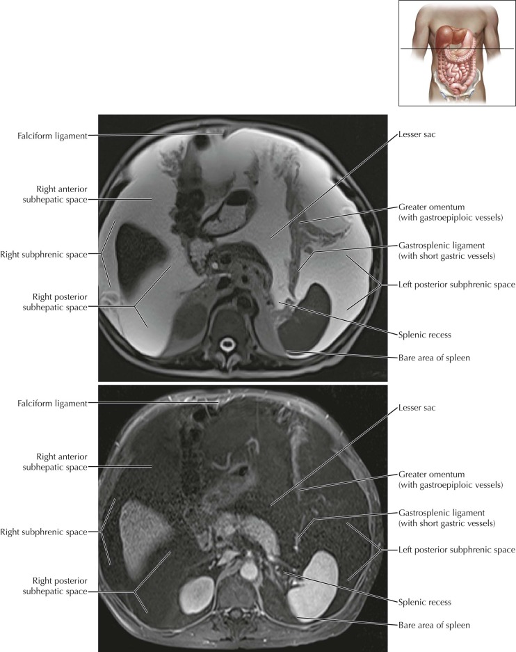
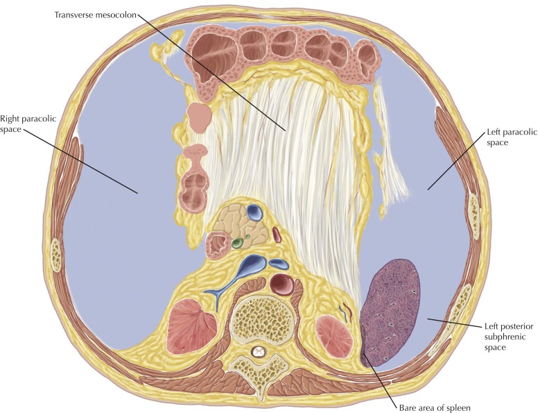
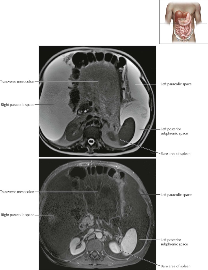
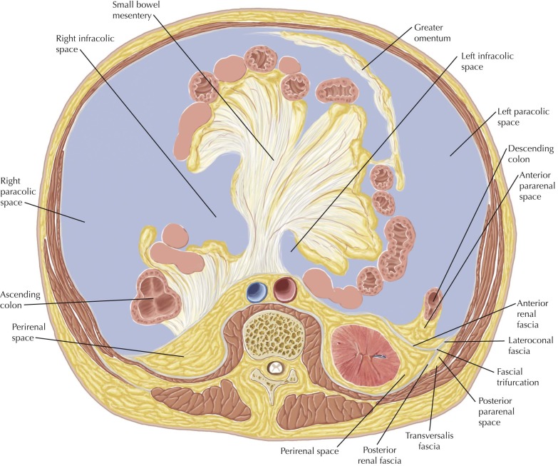
The abdominal peritoneal cavity can be divided by the transverse mesocolon into the supramesocolic and inframesocolic compartments. The supramesocolic compartment can be subdivided into the subphrenic spaces, subhepatic spaces, and lesser sac; the inframesocolic compartment can be subdivided into the infracolic and paracolic spaces. The greater sac of the abdominal peritoneal cavity comprises the subphrenic, subhepatic, infracolic, and paracolic spaces.
Pathologic Process
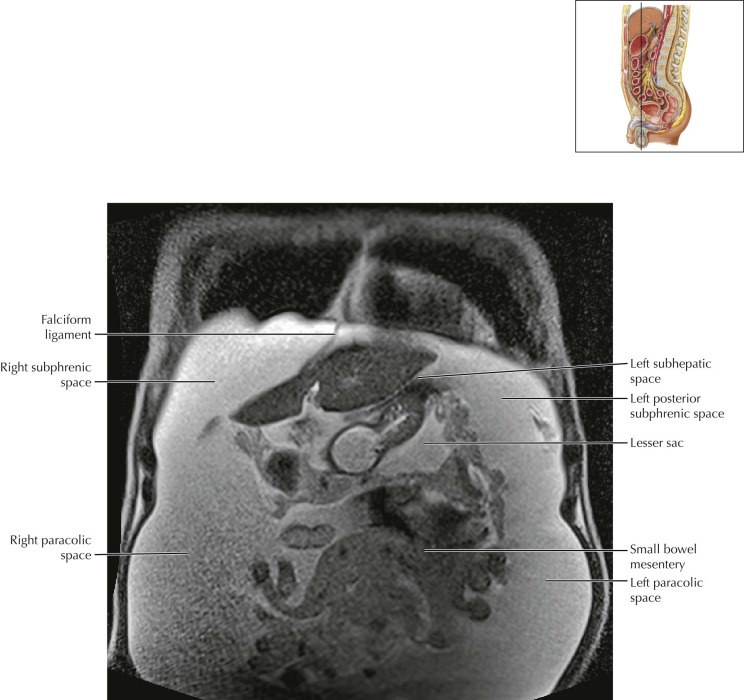
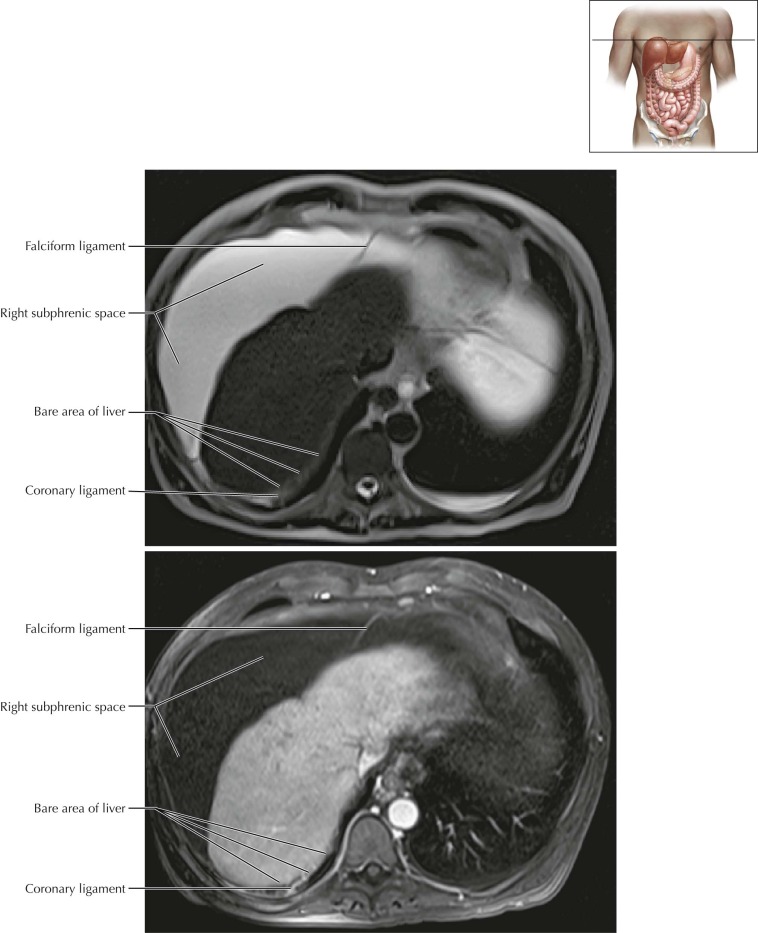
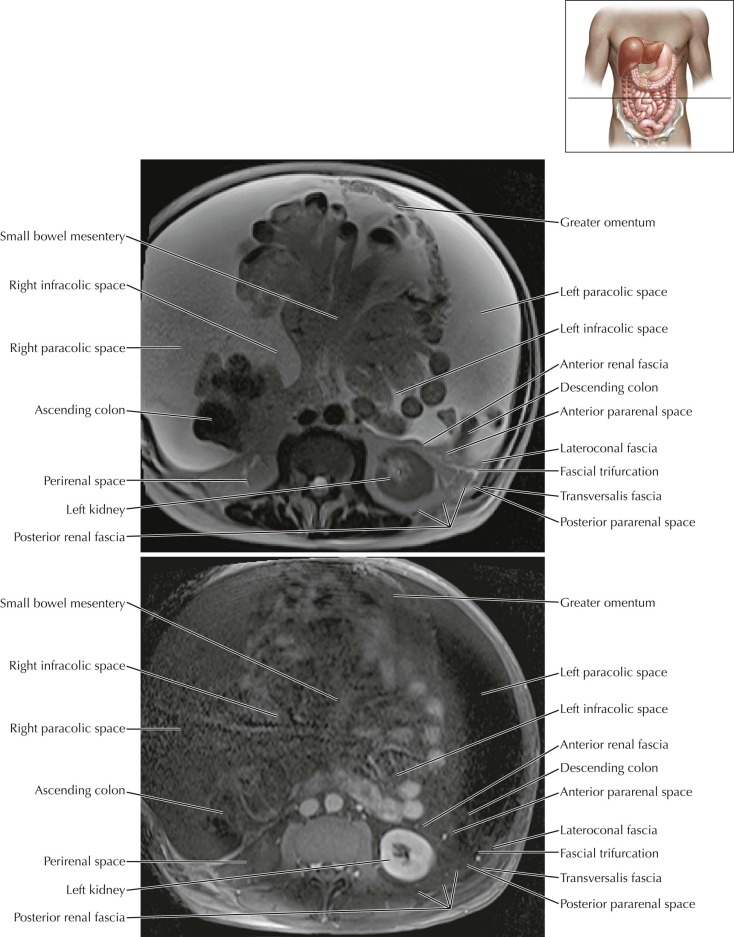
The spaces in the peritoneal cavity are best demonstrated in patients with ascites, which distends the potential space of the peritoneal cavity and outlines the peritoneal reflections, peritoneal ligaments, mesenteries, and omenta, as demonstrated in this example.
Peritoneal Cavity–Abdomen Axial 2
Normal Anatomy
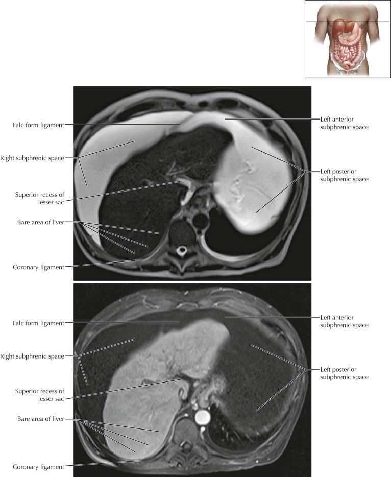
The right subphrenic space is bounded by the falciform ligament anteromedially and the right coronary ligament posteriorly. Posteromedial to the right coronary ligament is the bare area of the liver, the nonperitonealized attachment of the liver to the diaphragm, where peritoneal ascites is excluded. The bare area of the liver is continuous with the retroperitoneum.
Peritoneal Cavity–Abdomen Axial 3
Normal Anatomy
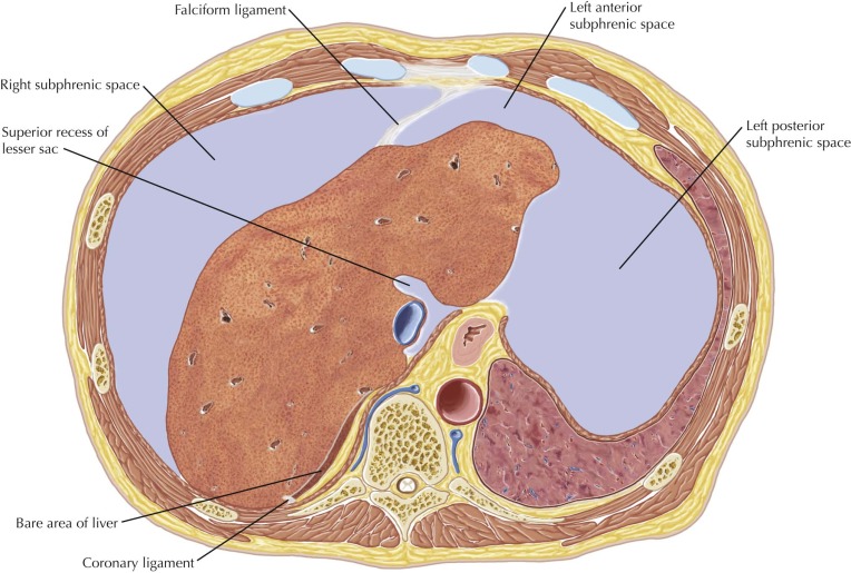
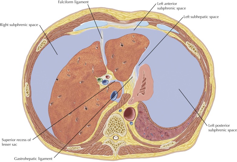
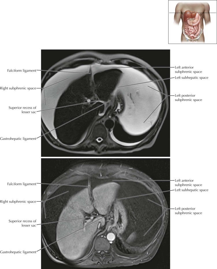
The left anterior subphrenic space extends from the falciform ligament anteromedially to surround the anterior left hepatic lobe and anterior gastric wall deep to the diaphragm. The inferior portion of this space, located between the left hepatic lobe posteriorly and the diaphragm anteriorly, may be considered a separate space, called the left anterior perihepatic space, which is bounded posteriorly by the left coronary ligament. The left posterior subphrenic space completely envelops the spleen and is also known as the “perisplenic space.”
The left subhepatic space, also known as the “left posterior perihepatic space” or “gastrohepatic recess,” is located between the lateral segment of the liver anteriorly and the stomach posteriorly, to the left of the gastrohepatic ligament.
The superior recess of the lesser sac is also seen at this level, adjacent to the caudate lobe of the liver (segment I).
Peritoneal Cavity–Abdomen Axial 4
Normal Anatomy
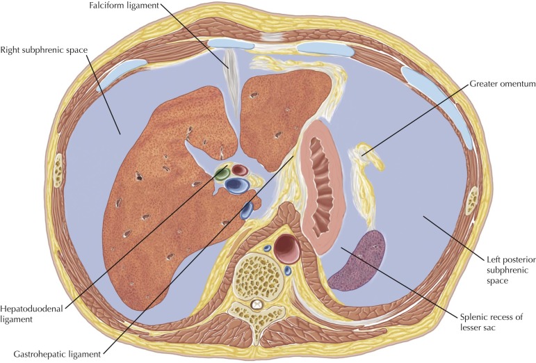
The hepatoduodenal and gastrohepatic ligaments form the lesser omentum (peritoneal fold) and can be seen at this level. The gastrohepatic ligament can be found by locating the left gastric artery, which runs within the ligament. The coronary vein also runs within the gastrohepatic ligament.
The splenic recess of the lesser sac is also seen at this level, between the gastric fundus and spleen.
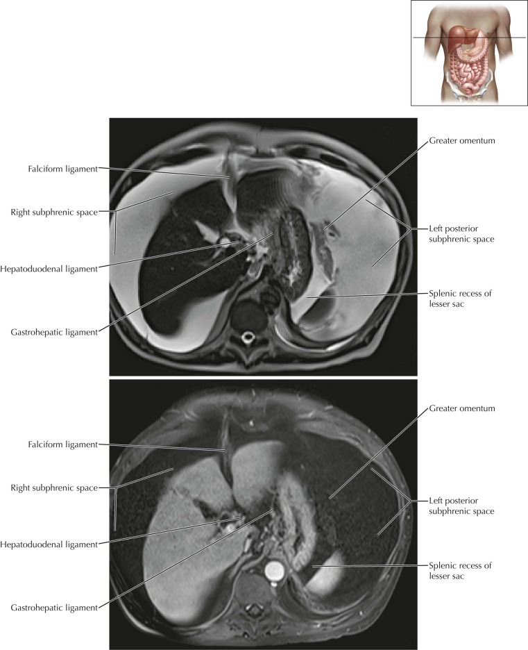
Peritoneal Cavity–Abdomen Axial 5
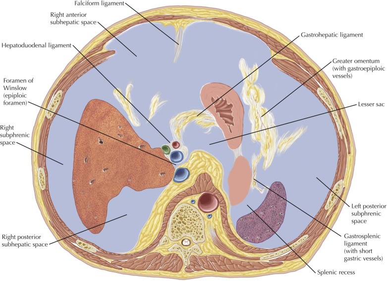
Normal Anatomy
The hepatoduodenal ligament extends from the porta hepatis to the duodenum and contains the proper hepatic artery, main portal vein, and common bile duct. The hepatoduodenal ligament forms the anterior margin of the foramen of Winslow, also known as the epiploic foramen, which is the communication between the lesser and greater sacs.
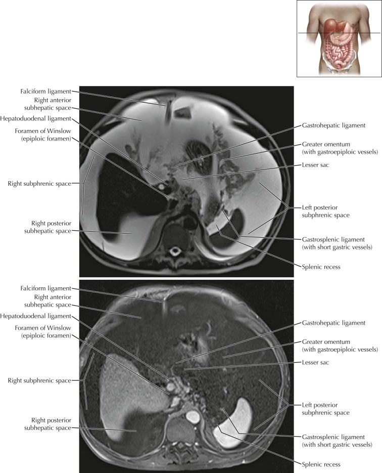
Peritoneal Cavity–Abdomen Axial 7
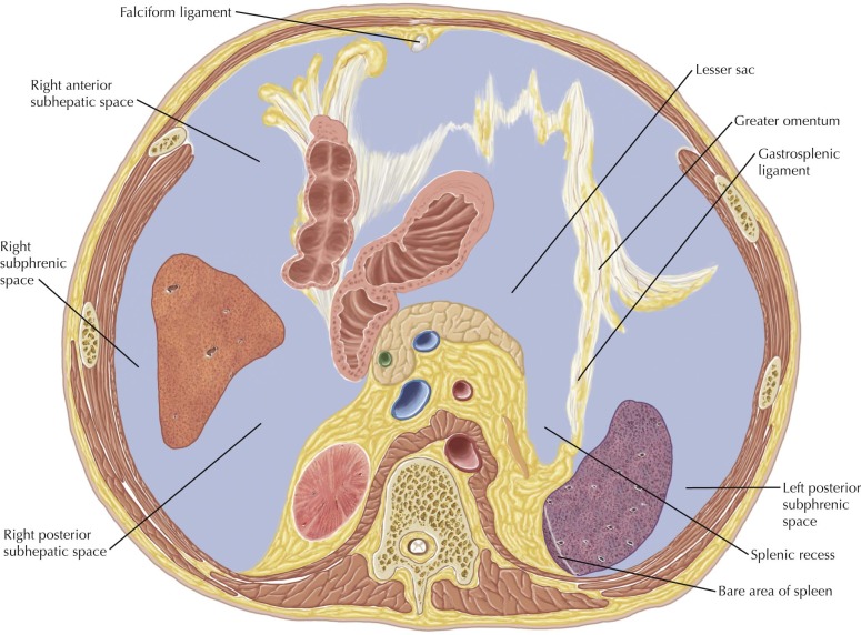
Normal Anatomy
The right subhepatic space (with anterior and posterior portions) is bounded superiorly by the inferior right lobe of the liver and is continuous with the right subphrenic space and right paracolic space. The right posterior subhepatic space, also known as Morison’s pouch or the hepatorenal space, is seen between the posterior right hepatic lobe and the right kidney, and is the most dependent portion of the abdominal peritoneal cavity in the supine position. As such, this space is a common location for peritoneal fluid to accumulate.
Peritoneal Cavity–Abdomen Axial 8
Normal Anatomy
The spleen is attached posteriorly to the retroperitoneum by the splenorenal ligament, forming the bare area of the spleen. The splenic artery and vein travel within the splenorenal ligament at the splenic hilum.
The transverse mesocolon is seen extending from the anterior aspect of the retroperitoneum at the level of the pancreas to the posterior superior wall of the transverse colon, dividing the abdominal peritoneal cavity into the supramesocolic and inframesocolic compartments described earlier. This may serve as a conduit for the spread of disease from the retroperitoneum to the transverse colon, as in the patient with acute pancreatitis.
Peritoneal Cavity–Abdomen Axial 10
Normal Anatomy
The retroperitoneum can be divided into three spaces. The anterior pararenal space is bounded anteriorly by the posterior parietal peritoneum and posteriorly by the anterior renal fascia (ARF), also called Gerota’s fascia, and contains the ascending and descending colon, pancreas, and the 2nd to 4th portions of the duodenum. The perirenal space is formed by the ARF, the lateroconal fascia, and the posterior renal fascia (PRF), also known as Zuckerkandl’s fascia, and contains the kidneys, adrenal glands, renal vasculature, and lymphatics, as well as the bridging renal septa of Kunin, which can serve as a conduit for disease spread through the perirenal space. The posterior pararenal space is bounded by the PRF and lateroconal fascia anteriorly and the transversalis fascia posteriorly, and is contiguous with the properitoneal fat anteriorly and laterally.
Peritoneal Cavity–Abdomen Coronal 2
Normal Anatomy
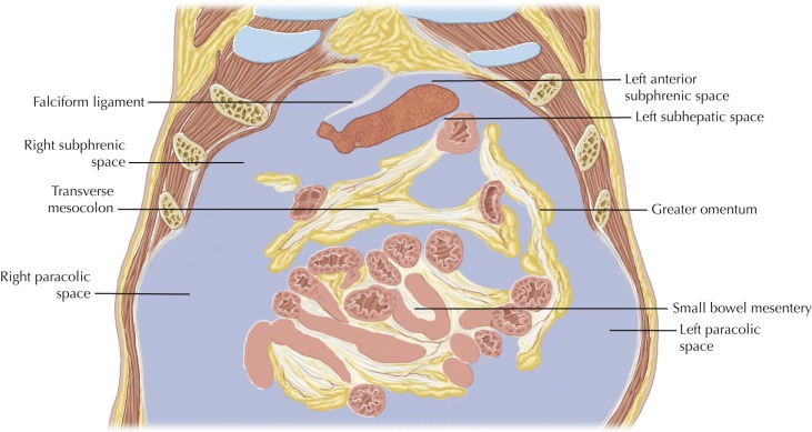
The superior and inferior portions of the peritoneal cavity communicate through the right and left paracolic spaces, also known as the “paracolic gutters,” which are formed by peritoneal reflections covering the colon and the abdominal wall laterally.
Peritoneal Cavity–Abdomen Coronal 3
Normal Anatomy
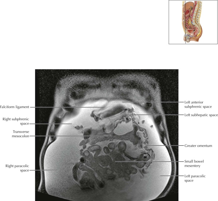
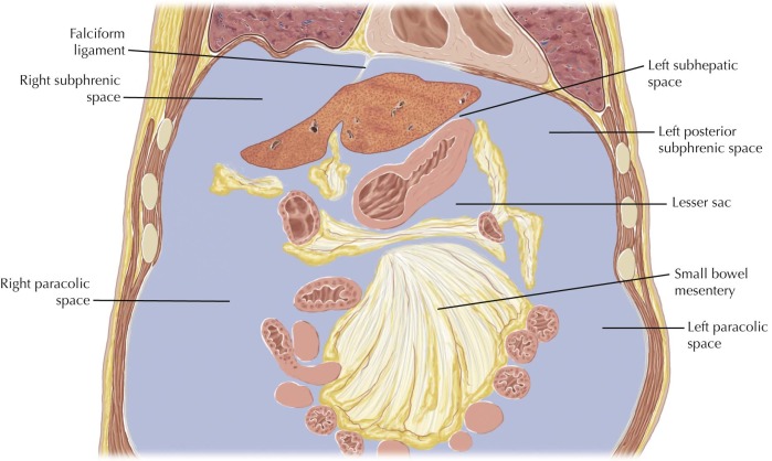
The right subphrenic space extends from the falciform ligament anteromedially to surround the diaphragmatic surface of the right lobe of the liver and is continuous with the right subhepatic space (seen on Abdomen Coronal 4 ) and the right paracolic space inferiorly. The right paracolic space communicates freely with the right pelvic peritoneal space.
The left subphrenic space extends from the falciform ligament anteromedially to surround the diaphragmatic surface of the left lobe of the liver and spleen, and is continuous with the left subhepatic space. The left subphrenic space is limited posteriorly and inferiorly by the splenorenal and phrenicocolic ligaments and more superiorly by the gastrosplenic ligament. Adjacent to the lateral segment of the liver, the left subhepatic space is continuous with the left subphrenic space.

