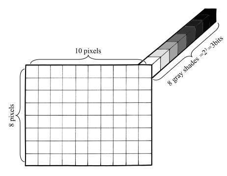
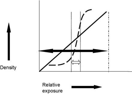
2 Physics of Digital Mammography A basic understanding of the principles of physics is useful in evaluating equipment and understanding the factors that affect the quality of the digital image. Digital signals are transmitted in multiples of defined equal signals. A common item that displays either analog or digital information is the watch. The second hand of the analog watch sweeps around the dial in a continuous 360-degree motion. One second is not discretely separated from the next. In fact, theoretically, it should be possible to time actions to a tenth or a hundredth of a second. However, practically speaking, the second hand moves too quickly for this type of measurement. When a digital watch displays seconds, the watch presents each second as a discrete whole number. The watch does not show any intervening tenth or hundredth of a second. There is only a change in the number when one whole second has passed. With digital transmission, information is relayed in discrete packets of information. Digital information is described using the terms pixels and bits or bytes. A pixel is a picture element. A digital image consists of a rectangular matrix of pixels. The size of the digital image is described as the number of horizontal pixels by the number of vertical pixels. Therefore, an image identified as 500 by 300 pixels refers to a rectangular matrix that is 500 pixels wide and 300 pixels high (Fig. 2–1). Because mammograms are displayed in various shades of gray, each pixel can potentially display a fixed maximal number of gray shades. The number of shades is described by bits. A bit is defined as 2X, where X is the number of bits. So, 8 bits is 256 shades of gray. A 12-bit image has 4096 shades of gray. Finally, 8 bits equals 1 byte. The total size of an image is described as the total number of pixels multiplied by the number of bits per pixel. Therefore, if the previously described image consisting of 500 by 300 pixels had a gray scale consisting of 8 bits, the total number of bits in the image would be 500 × 300 × 8 or 1,200,000 bits or 150,000 bytes. Familiarity with this numerical terminology is important because in digital imaging, these terms are used for a variety of applications: to express the amount of information within a digital mammogram, to describe the resolution of a workstation monitor, and to measure the amount of digital space needed for long-term storage.1 Figure 2–1 Schematic illustrating digital image matrix. The image consists of a matrix of picture elements (pixels). A digital image consists of a rectangular matrix of pixels. The size of the digital image is described by the number of horizontal pixels by the number of vertical pixels. Therefore, this rectangular matrix image is identified as 10 by 8 pixels. Each pixel is associated with a fixed number of gray-scale shades defined by bits (a bit is defined as 2X, where X is the number of bits). This image has eight shades of gray, or 23 or 3 bits. The total size of an image is described as the total number of pixels multiplied by the number of bits per pixel. Therefore, the total size of this image is 10 × 8 × 8 or 640 bits, or 80 bytes. One major difference in the physics of the digital mammography system and a screen-film system is the digital mammographic detector. The digital detector replaces the screen and film components of nondigital or analog mammography machines. The image produced by screen-film mammography is described by the characteristic curve that plots the relationship between film density on the vertical axis and the relative exposure on the horizontal axis (Fig. 2–2). In order to produce a mammogram that has a wide range of tissue densities, you should use exposures that result in densities that align with the steep part of the curve. This set of densities is the latitude of the film. Screen-film mammography has a relatively narrow latitude. However, digital detectors are created so that there is a linear relationship between density and relative exposure. This relationship produces a wider latitude. The latitude of digital mammography is ~1000:1 compared with 40:1 for screen film mammography. This wider latitude results in a broader range of exposures that will produce an acceptable image.2,3 Figure 2–2 Characteristic curves for digital detectors and film. The relationship between exposure (horizontal axis) and density (vertical axis) is shown by the black dashed curve for film and the solid slanted line for digital detectors. The narrow latitude (dotted arrow) of film is contrasted with the wide latitude (solid black arrow) for digital detectors.1 Digital latitude is related to dynamic range or the depth of recorded signal intensity. In a gray-scale image, dynamic range is recorded as a series of gray-scale shades. The numerical value of the dynamic range is expressed as bits or 2X steps of digitalization. The dynamic range required for digital mammographic imaging is related to the number of gray-scale shades that will adequately display the structures that attenuate the most and the fewest x-rays within the breast. The attenuation of x-rays is related to three factors: the energy of the x-ray, the breast thickness, and the breast tissue (fatty vs. fibroglandular). The following example demonstrates the general method that is used to determine the adequacy of digital mammographic detector dynamic range. In this example, to simplify the problem, you would make the following assumptions: (1) The incident x-ray is a monoenergetic 25 keV (kiloelectronvolt). (2) The breast is uniformly compressed to only 8 cm. (3) Part of this breast is composed of 0% fibroglandular tissue (i.e., almost entirely fat). (4) Part of this breast is composed of 100% fibroglandular tissue (this is extremely dense composition). After the incident x-ray passes through the hypothetical breast, the relative attenuation produced by the fibroglandular composition is calculated, taking into account the breast thickness and the x-ray keV. The calculated factors for this hypothetical mammogram are authentic and displayed in Table 2–1.4
 Image Acquisition Physics
Digital Detector Characteristic Curve Versus Screen-Film
Stay updated, free articles. Join our Telegram channel

Full access? Get Clinical Tree


