Location: (a) Paratendinous (frequent): in the sheath of a flexor tendon of the fingers, in the palm of the hand close to a metacarpophalangeal joint, in the wrist, on the dorsum of a finger adjacent to an extensor tendon, and rare in the foot; (b) in the joint (rare): >75 % in the knee, then in the hip, wrist, ankle, and shoulder; and (c) in the bursae (exceptional).
Clinical: Pain, swelling, effusion, or completely asymptomatic.
Diagnosis: On x-ray thickening of the synovial membrane with the same density of the soft tissue and without calcification. Skeletal erosions due to compression are frequent as rounded or multilobulated osteolytic lesions with well-defined margins, on the perimeter of the joint. Erosions are multiple and superficial, with sclerotic edges. On CT—lobulated newly formed tissue in the joint with considerable uptake of contrast dye. On bone scan—bone uptake and increased flow and blood pool in the mass. On MRI—heterogeneous, mostly low signal on T1 and T2 is characteristic. Intra- and peritumoral enhancing curvilinear regions on contrast T1.
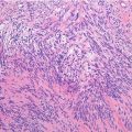
Histopathology: Roughly lobulated, single, soft, yellow-white to pale-brown nodules with smooth surface. In advanced stages it matures in a fibrous scar. It becomes hard, compact, and white with some yellow or brown bands and adheres to the surrounding tissues, to bone, and to tendon. The synovial membrane appears thickened, leathery yellow, and matted by long large villi like a “ruffled beard,” with multiple, soft, yellow-brown, lobulated nodules of varying size. Fibrin membranes cover the villi surface. Soft, pasty, friable, and yellow-brown tissue fills joint space in more advanced lesions. Pathologic tissue may be easily enucleated from the bone lesions that have a smooth bony wall. PVNS may invade the joint capsule and expand into the muscles, between the tendons, but it never infiltrates them.
Related posts:


Stay updated, free articles. Join our Telegram channel

Full access? Get Clinical Tree


