Fig. 2.1
Upright posteroanterior chest radiograph: evidence of free air beneath the right and left hemidiaphragms
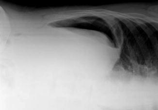
Fig. 2.2
Left lateral decubitus film of the abdomen in a patient with free peritoneal air resulting from gastric perforation
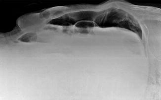
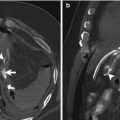
Fig. 2.3
Cross-table lateral abdominal radiograph demonstrating the presence of pneumoperitoneum
On supine abdominal radiograph, free peritoneal air may become visible and, in various shapes and sizes, may be located in different positions. These free air signs can be categorized into four groups: bowel-related signs, right upper quadrant signs, peritoneal ligament-related signs, and other signs [13].
Bowel-related signs include the following:
Rigler sign. The Rigler sign (Fig. 2.4), also known as the bas-relief sign or the double-wall sign, is the visualization of both sides of the bowel wall, on a radiograph of the abdomen obtained with the patient in the supine position, in the presence of a large volume of free air so that the bowel loops can be separated from each other [14].
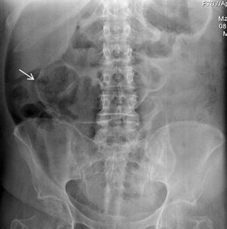
Fig. 2.4
Anteroposterior supine abdominal radiograph shows the Rigler sign (arrow)
Triangle sign. Free intraperitoneal air accumulating among three adjoining bowel loops or two bowel loops and the parietal peritoneum (Fig. 2.5) appearing as a triangular radiolucency is called the triangle sign [15].
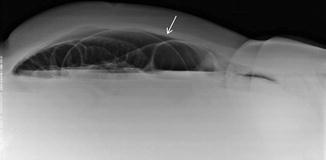
Fig. 2.5
Cross-table lateral radiograph of the abdomen showing the triangle sign (arrow)
Right upper quadrant signs include the following:
Hyperlucent liver sign. On the supine radiographs, the blacker density of the large intraperitoneal free gas anterior to the ventral hepatic surface replacing the brightness of the hepatic shadow is called the hyperlucent liver sign (Figs. 2.6 and 2.7) [15].
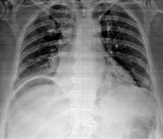
Fig. 2.6
Anteroposterior supine chest radiograph shows the hyperlucent liver sign
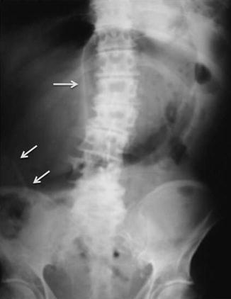
Fig. 2.7
A supine radiograph of the abdomen showing the hyperlucent liver sign, the falciform ligament sign (arrow), and the mesoappendix sign (arrows) in a perforated patient
Anterior superior oval sign. This sign refers to a single or multiple oval, round, or pear-shaped gas bubbles projected over the liver shadow [15, 16].
Fissure for ligamentum teres sign. This sign refers to a characteristic elongated area of hyperlucency that represents intraperitoneal gas trapped within the fissure for the ligamentum teres [17].
The visible gallbladder. On supine abdominal radiograph, the gallbladder is seen as a homogeneous opacity because of surrounding free intraperitoneal air [18].
Doge cap sign. This sign refers to triangle-shaped free air accumulated in Morison pouch on supine abdominal films [15, 19].
Hepatic edge sign. An oblong saucer or cigar-shaped collection of free air may be seen in the subhepatic space with its long axis directed superomedially following the liver contour [15, 20].
Dolphin sign. The undersurface of the long costal muscle slips of the diaphragm that indented the adjacent air-filled space in the right upper quadrant on supine films is a sign of pneumoperitoneum [21].
Peritoneal ligament-related signs include the following:
Falciform ligament sign. The intraperitoneal free air may outline the falciform ligament, which is seen as a linear density situated longitudinally within the right upper abdomen (Fig. 2.7) [15, 22].
Extrahepatic ligamentum teres sign. The ligamentum teres is another anterior peritoneal ligament that can be visualized on plain radiographs. It is a firm fibrous cord representing the remnant of an obliterated left umbilical vein. On supine radiographs, the extrahepatic ligamentum teres may be seen when outlined by free air anywhere along the course of the ligament [23].
The “inverted V” sign. Free air outlining the lateral umbilical ligaments makes these structures visible in the lower abdomen, forming an “inverted V” as they course inferiorly and laterally from the umbilicus [24, 25].
Urachus sign. When pneumoperitoneum occurs, the urachus may be seen as a thin midline linear structure in the lower abdomen from the umbilicus to the dome of the urinary bladder [26].
The transverse mesocolon and root of small bowel mesentery signs. The intraperitoneal free air can determine the identification of the transverse mesocolon and the root of the small bowel mesentery on plain abdominal radiographs obtained in the supine and in the prone position [27].
The mesoappendix sign. In the presence of a large amount of pneumoperitoneum, the mesoappendix may be observed on the supine radiograph as a radiopaque linear stripe directed from the cecum to the middle of the abdomen (Fig. 2.7).
Other signs of pneumoperitoneum are the following:
Football sign. It refers to a large oval radiolucency in the shape of an American football producing a sharp interface with the parietal peritoneum on a supine abdominal radiograph. The oval radiolucency seen in the football sign represents massive pneumoperitoneum which distends the peritoneal cavity [28, 29].
Cupola sign. The cupola sign is seen as an arcuate lucency overlying the lower thoracic spine and projecting caudad to the heart on supine radiograph [30]. The term cupola is used to indicate the inverted cup-shaped configuration of the lucency.Related posts:
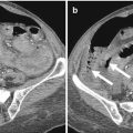
 Role of Multidetector Row Computed Tomography in the Diagnosis of Gastroduodenal Perforation
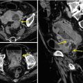
 MDCT Imaging of Gastrointestinal Tract Perforation Due to Foreign Body Ingestion
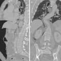
 Imaging of Gastrointestinal Tract Perforation in the Elderly Patient
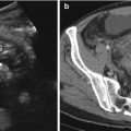 Ultrasonographic Assessment of Gastrointestinal Perforation
 Acute Perforated Diverticulitis: Spectrum of MDCT Findings
 Pneumoretroperitoneum: Imaging Findings

Stay updated, free articles. Join our Telegram channel

Full access? Get Clinical Tree


