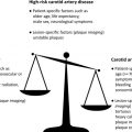
The continued occurrence of stroke despite advances in medical therapy for asymptomatic carotid stenosis (ACS) strongly indicates that individual response to medical therapy may vary widely. This article reviews the literature that identifies MR imaging and ultrasound plaque features which are seen in patients at increased risk of future cardiovascular events. Imaging can identify plaque phenotype that is the most amendable to intensive medical therapy. There is also good evidence that plaque imaging can measure the individual response to medical therapy and the lack of response identifies a high-risk group of ACS patients.
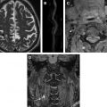
The presence of carotid intraplaque hemorrhage and/or ulceration in patients with asymptomatic carotid stenosis may require close monitoring to identify progression despite intensive medical therapy that is better treated with surgical intervention.
The presence of carotid intraplaque hemorrhage and/or ulceration in patients with asymptomatic carotid stenosis may require close monitoring to identify progression despite intensive medical therapy that is better treated with surgical intervention.
References
- 1. Bonita R., Stewart A., and Beaglehole R.: International trends in stroke mortality: 1970-1985. Stroke 1990; 21: pp. 989-992
- 2. Amarenco P., and Labreuche J.: Lipid management in the prevention of stroke: review and updated meta-analysis of statins for stroke prevention. Lancet Neurol 2009; 8: pp. 453-463
- 3. Stone N.J., Robinson J.G., Lichtenstein A.H., et al: 2013 ACC/AHA guideline on the treatment of blood cholesterol to reduce atherosclerotic cardiovascular risk in adults: a report of the American College of Cardiology/American Heart Association Task Force on Practice Guidelines. Circulation 2014; 129: pp. S1-S45
- 4. Kakkos S.K., Griffin M.B., Nicolaides A.N., et al: The size of juxtaluminal hypoechoic area in ultrasound images of asymptomatic carotid plaques predicts the occurrence of stroke. J Vasc Surg 2013; 57: pp. 609-618.e1
- 5. Paraskevas K.I., Spence J.D., Veith F.J., et al: Identifying which patients with asymptomatic carotid stenosis could benefit from intervention. Stroke J Cereb Circ 2014; 45: pp. 3720-3724
- 6. Markus H.S., King A., Shipley M., et al: Asymptomatic embolisation for prediction of stroke in the Asymptomatic Carotid Emboli Study (ACES): a prospective observational study. Lancet Neurol 2010; 9: pp. 663-671
- 7. Spence J.D., Coates V., Li H., et al: Effects of intensive medical therapy on microemboli and cardiovascular risk in asymptomatic carotid stenosis. Arch Neurol 2010; 67: pp. 180-186
- 8. Spence J.D., and Hackam D.G.: Treating arteries instead of risk factors: a paradigm change in management of atherosclerosis. Stroke J Cereb Circ 2010; 41: pp. 1193-1199
- 9. Takaya N., Yuan C., Chu B., et al: Association between carotid plaque characteristics and subsequent ischemic cerebrovascular events: a prospective assessment with MRI–initial results. Stroke 2006; 37: pp. 818-823
- 10. Xu D., Hippe D.S., Underhill H.R., et al: Prediction of high-risk plaque development and plaque progression with the carotid atherosclerosis score. JACC Cardiovasc Imaging 2014; 7: pp. 366-373
- 11. Saam T., Hetterich H., Hoffmann V., et al: Meta-analysis and systematic review of the predictive value of carotid plaque hemorrhage on cerebrovascular events by magnetic resonance imaging. J Am Coll Cardiol 2013; 62: pp. 1081-1091
- 12. Virmani R., Burke A.P., Kolodgie F.D., et al: Vulnerable plaque: the pathology of unstable coronary lesions. J Interv Cardiol 2002; 15: pp. 439-446
- 13. Naghavi M., Libby P., Falk E., et al: From vulnerable plaque to vulnerable patient: a call for new definitions and risk assessment strategies: Part I. Circulation 2003; 108: pp. 1664-1672
- 14. Naghavi M., Libby P., Falk E., et al: From vulnerable plaque to vulnerable patient: a call for new definitions and risk assessment strategies: Part II. Circulation 2003; 108: pp. 1772-1778
- 15. Stary H.C., Chandler A.B., Dinsmore R.E., et al: A definition of advanced types of atherosclerotic lesions and a histological classification of atherosclerosis. A report from the Committee on Vascular Lesions of the Council on Arteriosclerosis, American Heart Association. Circulation 1995; 92: pp. 1355-1374
- 16. Cai J.M., Hatsukami T.S., Ferguson M.S., et al: Classification of human carotid atherosclerotic lesions with in vivo multicontrast magnetic resonance imaging. Circulation 2002; 106: pp. 1368-1373
- 17. Spence J.D., and Parraga G.: Three-dimensional ultrasound of carotid plaque. Neuroimag Clin N Am 2015; undefined:
- 18. Underhill H.R., and Yuan C.: Carotid MRI: a tool for monitoring individual response to cardiovascular therapy? Expert Rev Cardiovasc Ther 2011; 9: pp. 63-80
- 19. Saam T., Kerwin W.S., Chu B., et al: Sample size calculation for clinical trials using magnetic resonance imaging for the quantitative assessment of carotid atherosclerosis. J Cardiovasc Magn Reson 2005; 7: pp. 799-808
- 20. Li F., Yarnykh V.L., Hatsukami T.S., et al: Scan-rescan reproducibility of carotid atherosclerotic plaque morphology and tissue composition measurements using multicontrast MRI at 3T. J Magn Reson Imaging 2010; 31: pp. 168-176
- 21. Underhill H.R., Yarnykh V.L., Hatsukami T.S., et al: Carotid plaque morphology and composition: initial comparison between 1.5- and 3.0-T magnetic field strengths. Radiology 2008; 248: pp. 550-560
- 22. Chu B., Kampschulte A., Ferguson M.S., et al: Hemorrhage in the atherosclerotic carotid plaque: a high-resolution MRI study. Stroke 2004; 35: pp. 1079-1084
- 23. Saam T., Ferguson M.S., Yarnykh V.L., et al: Quantitative evaluation of carotid plaque composition by in vivo MRI. Arterioscler Thromb Vasc Biol 2005; 25: pp. 234-239
- 24. Kampschulte A., Ferguson M.S., Kerwin W.S., et al: Differentiation of intraplaque versus juxtaluminal hemorrhage/thrombus in advanced human carotid atherosclerotic lesions by in vivo magnetic resonance imaging. Circulation 2004; 110: pp. 3239-3244
- 25. Ota H., Yarnykh V.L., Ferguson M.S., et al: Carotid intraplaque hemorrhage imaging at 3.0-T MR imaging: comparison of the diagnostic performance of three T1-weighted sequences 1. Radiology 2010; 254: pp. 551-562
- 26. Cai J., Hatsukami T.S., Ferguson M.S., et al: In vivo quantitative measurement of intact fibrous cap and lipid-rich necrotic core size in atherosclerotic carotid plaque: comparison of high-resolution, contrast-enhanced magnetic resonance imaging and histology. Circulation 2005; 112: pp. 3437-3444
- 27. Yuan C., Kerwin W.S., Ferguson M.S., et al: Contrast-enhanced high resolution MRI for atherosclerotic carotid artery tissue characterization. J Magn Reson Imaging 2002; 15: pp. 62-67
- 28. Narumi S., Sasaki M., Natori T., et al: Carotid plaque characterization using 3D T1-weighted MR imaging with histopathologic validation: a comparison with 2D technique. AJNR Am J Neuroradiol 2015; 36: pp. 751-756
- 29. Cheng J., Li H., Xiao F., et al: Fully automatic plaque segmentation in 3-D carotid ultrasound images. Ultrasound Med Biol 2013; 39: pp. 2431-2446
- 30. Græbe M., Entrekin R., Collet-Billon A., et al: Reproducibility of two 3-D ultrasound carotid plaque quantification methods. Ultrasound Med Biol 2014; 40: pp. 1641-1649
- 31. Schminke U., Motsch L., Hilker L., et al: Three-dimensional ultrasound observation of carotid artery plaque ulceration. Stroke J Cereb Circ 2000; 31: pp. 1651-1655
- 32. Madani A., Beletsky V., Tamayo A., et al: High-risk asymptomatic carotid stenosis: ulceration on 3D ultrasound vs TCD microemboli. Neurology 2011; 77: pp. 744-750
- 33. Sztajzel R., Momjian S., Momjian-Mayor I., et al: Stratified gray-scale median analysis and color mapping of the carotid plaque: correlation with endarterectomy specimen histology of 28 patients. Stroke 2005; 36: pp. 741-745
- 34. Griffin M.B., Kyriacou E., Pattichis C., et al: Juxtaluminal hypoechoic area in ultrasonic images of carotid plaques and hemispheric symptoms. J Vasc Surg 2010; 52: pp. 69-76
- 35. Markus H.S., and Molloy J.: Use of a decibel threshold in detecting Doppler embolic signals. Stroke J Cereb Circ 1997; 28: pp. 692-695
- 36. Ringelstein E.B., Droste D.W., Babikian V.L., et al: Consensus on microembolus detection by TCD. International Consensus Group on Microembolus Detection. Stroke J Cereb Circ 1998; 29: pp. 725-729
- 37. Kolodgie F.D., Gold H.K., Burke A.P., et al: Intraplaque hemorrhage and progression of coronary atheroma. N Engl J Med 2003; 349: pp. 2316-2325
- 38. Takaya N., Yuan C., Chu B., et al: Presence of intraplaque hemorrhage stimulates progression of carotid atherosclerotic plaques: a high-resolution magnetic resonance imaging study. Circulation 2005; 111: pp. 2768-2775
- 39. Moody A.R., Murphy R.E., Morgan P.S., et al: Characterization of complicated carotid plaque with magnetic resonance direct thrombus imaging in patients with cerebral ischemia. Circulation 2003; 107: pp. 3047-3052
- 40. Gupta A., Baradaran H., Schweitzer A.D., et al: Carotid plaque MRI and stroke risk: a systematic review and meta-analysis. Stroke J Cereb Circ 2013; 44: pp. 3071-3077
- 41. Underhill H.R., Hatsukami T.S., Cai J., et al: A noninvasive imaging approach to assess plaque severity: the carotid atherosclerosis score. AJNR Am J Neuroradiol 2010; 31: pp. 1068-1075
- 42. Spence J.D., Eliasziw M., DiCicco M., et al: Carotid plaque area: a tool for targeting and evaluating vascular preventive therapy. Stroke J Cereb Circ 2002; 33: pp. 2916-2922
- 43. Spence J.D.: Time course of atherosclerosis regression. Atherosclerosis 2014; 235: pp. 347-348
- 44. Spence J.D., Tamayo A., Lownie S.P., et al: Absence of microemboli on transcranial Doppler identifies low-risk patients with asymptomatic carotid stenosis. Stroke J Cereb Circ 2005; 36: pp. 2373-2378
- 45. Kuk M., Wannarong T., Beletsky V., et al: Volume of carotid artery ulceration as a predictor of cardiovascular events. Stroke J Cereb Circ 2014; 45: pp. 1437-1441
- 46. Van Engelen A., Wannarong T., Parraga G., et al: Three-dimensional carotid ultrasound plaque texture predicts vascular events. Stroke J Cereb Circ 2014; 45: pp. 2695-2701
- 47. Demarco J.K., and Huston J.: Imaging of high-risk carotid artery plaques: current status and future directions. Neurosurg Focus 2014; 36: pp. E1
- 48. Demarco J.K., Ota H., Underhill H.R., et al: MR carotid plaque imaging and contrast-enhanced MR angiography identifies lesions associated with recent ipsilateral thromboembolic symptoms: an in vivo study at 3T. AJNR Am J Neuroradiol 2010; 31: pp. 1395-1402
- 49. Hellings W.E., Peeters W., Moll F.L., et al: Composition of carotid atherosclerotic plaque is associated with cardiovascular outcome: a prognostic study. Circulation 2010; 121: pp. 1941-1950
Related posts:
 Three-Dimensional Carotid Plaque MR Imaging
Incorporating Carotid Plaque Imaging into Routine Clinical Carotid Magnetic Resonance Angiography
FDG PET/CT Imaging of Carotid Atherosclerosis
Clinical Perspective of Carotid Plaque Imaging
Three-Dimensional Carotid Plaque MR Imaging
Incorporating Carotid Plaque Imaging into Routine Clinical Carotid Magnetic Resonance Angiography
FDG PET/CT Imaging of Carotid Atherosclerosis
Clinical Perspective of Carotid Plaque Imaging
 Plaque Imaging to Decide on Optimal Treatment
 Low-Grade Carotid Stenosis
Stay updated, free articles. Join our Telegram channel

Full access? Get Clinical Tree


