and Vincent L. Sorrell2
(1)
Division of Nuclear Medicine and Molecular Imaging Department of Radiology, University of Kentucky, Lexington, Kentucky, USA
(2)
Division of Cardiovascular Medicine Department of Internal Medicine Gill Heart Institute, University of Kentucky, Lexington, Kentucky, USA
Electronic supplementary material
The online version of this chapter (doi: 10.1007/978-3-319-25436-4_9) contains supplementary material, which is available to authorized users.
The differential diagnosis of pleural findings on MPI is a short list (Table 9.1). Malignant lesions of the pleura can appear “hot” and are uncommonly identified on MPI (Chin et al. 1995; Eftekhari and Gholamrezanezhad 2006). A lung tumor might be associated with a “hot” or “cold” pleural effusion (Eftekhari and Gholamrezanezhad 2006); this topic is covered in more detail in Chap. 10 Lungs.
Table 9.1
Differential diagnosis of “hot” and “cold” imaging findings related to the pleura
Organ system | “Hot” finding | “Cold” finding | References |
|---|---|---|---|
Pleura | Neoplasm, malignant/mesothelioma Pleural effusion, malignant | Pleural effusion, benign Pleural effusion, malignant | Chamarthy and Travin (2010) Chin et al. (1995) Eftekhari and Gholamrezanezhad (2006) Gedik et al. (2007) Joy et al. (2007) Shih et al. (2002) Williams et al. (2003) |
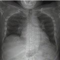
Pleural effusions, whether benign or malignant, are usually “cold” and are readily identified on MPI raw projection data (Chamarthy and Travin 2010; Gedik et al. 2007). They may be unilateral or bilateral and vary in volume from small to quite large (Shih et al. 2002). For SPECT MPI, the patient is positioned supine or semirecumbent, depending on the gamma camera system; and free pleural fluid will settle posteriorly. Right-sided pleural effusions are more difficult to appreciate unless they are large (Figs. 9.1 and 9.2). Left-sided pleural effusions are more readily apparent because the field-of-view extends more posteriorly around the left hemithorax, providing a more complete survey (Figs. 9.3 and 9.4). Left pleural effusions may be problematic because they may create attenuation artifact in the processed cardiac images with the inferior wall typically affected. Readers need to be aware of this potential source of artifact at the time of image interpretation. It is a good practice to access correlative imaging whenever available (Figs. 9.1, 9.2, 9.3, and 9.4) (Williams et al. 2003). Pleural effusions may be associated with abdominal ascites (Figs. 9.1, 9.3, and 9.4); this topic is covered in Chap. 18 Peritoneum (Joy et al. 2007).


Fig. 9.1


“Cold” right pleural effusion confirmed by other imaging. There is a “cold” region in the right lower hemithorax (a, b) that corresponds to a pleural effusion radiographically (c, d). There is no left pleural effusion (c, d). An isolated right pleural effusion typically does not affect the processed SPECT images; however, it may be clinically significant. Note a relative elevation of the left hemidiaphragm with “cold” ascites beneath it (a, e, f).(a) Stress raw projection images (Video 9.1a, frame 1), 99mTc sestamibi. (b) Stress raw projection image (Video 9.1b, frame 3), 99mTc sestamibi, “cold” pleural fluid (blue box), “cold” abdominal ascites (orange box). (c) Frontal chest radiograph, right pleural fluid (blue box). (d) CT at level of lower chest, right pleural fluid (blue box). (e) Stress raw projection image (Video 9.1b, frame 48), 99mTc sestamibi, “cold” ascites outlining the elevated left hemidiaphragm (orange lines). (f) CT at the level of upper abdomen, perihepatic and perisplenic ascites (orange boxes)
Stay updated, free articles. Join our Telegram channel

Full access? Get Clinical Tree


