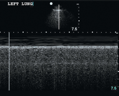
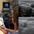
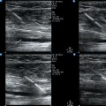
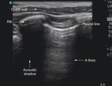
20 Proper equipment setup is important to maximize diagnostic yield, increase image detail, and improve accuracy. Usually, two-dimensional imaging is adequate for a pleural sonographic examination, and color Doppler techniques are not generally required. A microconvex transducer (3.5 to 8 MHz) is ideal for pleural ultrasound examination because it allows the deeper structures to be visualized and has a small footprint such that it can easily fit between the intercostal spaces.1 In addition, high-frequency transducers can be used to allow more detailed imaging of the pleural surface. In addition to transducer selection, proper orientation of the transducer is important when imaging the pleura and pulmonary structures. Imaging of the pleura should be performed in the longitudinal plane, and the marker of the transducer should always be directed to the head of the patient. By standard convention, imaging of the pleura will require the marker to be in the top left corner of the ultrasound screen.2 The operator is able to perform various adjustments to optimize the quality of acquired images as detailed in previous chapters. When performing a basic lung ultrasound examination, the thorax is usually divided in three zones: • The anterior zone is defined medially by the sternum, superiorly by the clavicle, laterally by the anterior axillary line, and inferiorly by the diaphragm. • The posterior zone extends from the posterior axillary line to the posterior midline. • The lateral zone is located between the anterior and posterior zones. The ribs are easily identified because they cast an acoustic shadow. The examiner should move the transducer on a longitudinal plane to identify the pleura and lung tissue through the intercostal spaces while scrolling through the ribs (Figure 20-1).2 Figure 20-1 Normal pleural line. A longitudinal view of the chest wall displays normal anatomic boundaries. The pleura appear as a moving bright line on lung ultrasonography. When using a high-frequency transducer the visceral and parietal pleura can be visualized as separate linear structures that are closely apposed. Movement of the pleural line against a fixed chest wall is known as lung sliding. Visualization of lung sliding implies that the two pleural surfaces are mobile and in apposition with each other and that no air is opposed between them at the point of the transducer. It is very important to distinguish pleural sliding from chest wall movement.1,2 An alternative method of assessing lung sliding is M-mode. Pleural sliding gives a characteristic seashore sign (Figure 20-2). The presence of lung sliding implies with 100% certainty that no pneumothorax is present at the point of the probe.3 Careful observation of the pleura reveals a subtle shimmering movement of the pleural line that coincides with cardiac pulsation; this is known as lung pulse. The finding of lung pulse is usually more prominent in the left hemithorax and in the lower aspects of the thorax.2 The presence of lung pulse indicates apposition of the parietal and visceral pleural surfaces at the point of the transducer; therefore the finding of lung pulse reliably excludes the presence of pneumothorax. The ability to rule out pneumothorax quickly is a critical application of pleural ultrasonography, which is noninvasive procedure that has greater specificity than and equal sensitivity as chest radiography in diagnosing pneumothorax.4 It is also excellent for detecting residual pneumothorax after drainage.5 At the point on the chest where the pneumothorax is present there will be neither lung sliding and nor a seashore sign. Because the air rises to nondependent areas in the pleural cavity, ultrasound examination should begin in this area of the chest.2 The presence of lung sliding rules out pneumothorax. However, the opposite is not true, and the examiner should keep in mind that the absence of lung sliding is not specific for pneumothorax only. Lung sliding may be absent in other conditions such as atelectasis, pleural thickening, extensive pulmonary fibrosis, and pleural symphysis.1 Other findings that suggest the presence of pneumothorax are lung point, “barcode” sign (Figure 20-3), and B-lines (lung rockets), as detailed in Chapter 19.2,
Pleural ultrasound
Overview
Identification of anatomic structures
Lung sliding
Lung pulse
Pneumothorax
![]()
Stay updated, free articles. Join our Telegram channel

Full access? Get Clinical Tree


Radiology Key
Fastest Radiology Insight Engine

