Point-of-Care


Point-of-Care


Javier Rosario


Point-of-care (POC) sonography—or, more commonly, point-of-care ultrasonography (POCUS)—can be characterized into five fundamental clinical categories: diagnostic, monitoring, procedural guidance, resuscitation, and symptom- or sign-based. Using POCUS for diagnostic imaging allows the clinician to focus on distinctive organ pathologies based on the patient’s presenting signs and symptoms. Having the ability to quickly distinguish between normal and abnormal anatomical findings allows for tapering of differential diagnoses and determining the best management strategies, while still at the bedside or in the immediate clinical setting. Focused ultrasonographic examinations can be performed on any organ or cavity that full diagnostic sonography currently excels at. Monitoring of certain conditions via POCUS allows for specific serial sonographic assessments of a patient’s known condition or to observe the effects of an ongoing intervention. The use of ultrasound for procedural guidance has long been proven to reduce complications and enhance success rates because of its ability to dynamically and in real time visualize and track the procedural process. The adoption of POCUS in the trauma setting specifically during resuscitation and evaluation of traumatic injuries during cardiac arrest has been widely accepted and is already a well-established application within the guidelines of traumatic evaluation. The ability to immediately assess and direct emergent interventions by swiftly diagnosing tension pneumothorax, cardiac tamponade, and massive intraperitoneal fluid is well appreciated within the literature.1, 2 and 3 Skilled clinicians may be able to determine cardiac standstill, clotting within ventricular and atrial chambers, or visualization of subtle contractions or fibrillation during cardiac arrest and appropriately guide care. A symptom- or sign-based evaluation carries many advantages because of its ability to rapidly assess patients with their given signs and symptoms and may help further improve the diagnostic accuracy of the differential diagnosis while also offering the avoidance of ionizing radiation. Today, many subspecialties are using POCUS as a principal tool to their daily practices. The term sonologist may be interchanged or used when clinicians performing these studies are making direct clinical determinations based on their findings.

Throughout the last decade, ultrasound imaging and information systems have continued decreasing in size, while at the same time becoming more sophisticated and increasing their usability. These advances have increased the versatility, mobility, and integration of a broad spectrum of clinical applications in which they can be used. For this reason, sonography is far from being limited to the traditional radiology or cardiology settings. Sonography continues to establish itself as an indispensable tool in the evaluation of acute patients in many realms within the prehospital, hospital, and in the field/office setting. The technological advancements in instrumentation allow for a less complicated user interface, which increases the ease of operating the equipment and allows for its application in multiple health care settings.


POCUS has been described as sonographic imaging used to enhance and advance patient care during a specific encounter or procedure. Today, POCUS covers a broad spectrum of medical specialties in a variety of settings because it has been shown to improve the safety and effectiveness of patient care.4, 5 and 6 Most clinical applications involve answering a specific question, or set of questions, through a focused and directed sonographic examination to better assess if a condition or pathology is present or the cause of the patient’s symptoms.

For several decades, the use of ultrasound evaluations for patient care has become an integral part of specialties like emergency medicine (EM) and acute and critical care. Recently, there has been increasing interest on the utility of POCUS for patient care in almost every medical and surgical specialty.7 Now, POCUS is used to evaluate most body systems and virtually every disease process with great effectiveness (Table 25-1).

This chapter focuses on some of the primary indications for sonographic examination in the POC setting and discusses how sonography is more readily utilized on both acute and stable patients in diverse areas of clinical practice.


PERFORMANCE STANDARDS

Sonography is a well-established, reliable, and noninvasive diagnostic tool. This concept of quick and limited sonographic imaging began spreading in Japan and Europe in the 1970s.2 However, it was not until the 1980s and 1990s that physicians in the United States began publishing studies focused on the depiction of free fluid or blood within the peritoneal and pericardial spaces to aid with critical trauma settings. Since that time, the use of targeted sonographic assessments has evolved and has been utilized in multiple specialties within medicine. As the medical profession continues to gain experience with sonography and as the technology continues to ascend, its varied use in more clinical applications and in a broader spectrum of disciplines is continually growing as well.8, 9 and 10

The growth in the applications of these sonographic assessments has led to an evolving standard of care developed from what began as best practice for emergency department trauma care to now being included in inpatient units and
outpatient settings.11,12 As with any growth, there are continued challenges to ensure whether its full potential and diagnostic abilities are being reached. These include but are not limited to adequate training, proper documentation and image archiving for all to adhere to, meeting standard competencies, and proper governance or oversight. Once these important criteria have been established and fulfilled, proper continuing education requirements should be met, and the necessary skill sets should continually be developed. Another important factor to successful POCUS adaptation is the utilization of proper equipment. The lack of proper equipment may limit the type of applications available for patient care and the thoroughness of its adoption.


Professional associations have developed recognized guidelines, recommendations, and standardizations for bedside sonographic examinations because POCUS has branched to become an extra set of skills that can enhance the traditional clinical examination. Two well-known associations that have shown strong support to the use of POCUS with websites containing valuable clinical information include the American College of Emergency Physicians and the American Institute of Ultrasound in Medicine.13,14 A brief summary of the practice guidelines for emergency sonography published by these organizations is given in Table 25-2. Each publication includes standards for personnel, education, protocols, risk management, quality control, quality improvement, and scanning equipment and maintenance.


CLINICAL APPLICATIONS


Emergency Medicine

For several decades, the specialty of EM has established itself as a leader in the education and advancement of POCUS in the clinical setting. Emergency physicians receive training in POCUS since their early training in residency and have established robust guidelines for its use in the clinical setting (Table 25-3).13 For advanced and expanded applications, there are over 120 EM-focused fellowship programs providing advanced clinical ultrasound training under standardized curricula and guidelines.15 Whereas most of these guidelines are EM-specific, other medical professionals have taken interest in POCUS applications and use these to formulate their own individual and specialty-specific guidelines. The following sections will further break down these POCUS core applications. However, to obtain more expanded evaluations of specific organ systems, other chapters of this book may be recommended.


Trauma and Extended Focused Assessment with Sonography in Trauma Examination

The focused assessment with sonography in trauma (FAST) examination originated in the United States in the early 1990s and was likely the original POC rapid sonographic examination.16,17 The FAST examination and the extended FAST (eFAST) examination are two protocols used to detect sequelae of trauma in emergency sonography. Implementing the eFAST for emergency sonography provides a rapid evaluation tool for the everyday practice of trauma and/or acute patient care, which can significantly improve patient care and triage and balance the differences between medical and surgical emergencies. Following the advanced trauma life support (ATLS) guidelines, it can be utilized in both stable and unstable patients, in conjunction with the physical examinations, resuscitation, and stabilization. The concept behind the FAST examination is based on the fact that many life-threatening injuries cause bleeding in the pericardium, thorax, abdomen, and pelvic regions. The primary purpose of the FAST examination is the methodical search for anechoic free fluid (i.e., blood in the appropriate setting) in the dependent regions of the pericardium, pleural spaces, intraperitoneal spaces, and the retroperitoneum. The primary purpose of the eFAST examination is to extend the search for a pneumothorax in this same scenario.

The eFAST examination can be up to 96% specific for detecting any amount of intraperitoneal free fluid18 and nearly perfect for the detection of intraperitoneal bleeding significant enough to cause shock and an emergent laparotomy.19, 20, 21, 22 and 23 The use of the eFAST examination has made the use of diagnostic peritoneal lavage (DPL) obsolete in the acute evaluation of traumatic injuries. It is important to understand that the eFAST examination does not provide a comprehensive examination of the underlying pathology. However, there are major benefits to performing this
noninvasive bedside examination, which can be performed on pregnant women and children, without the risk of exposure to ionizing radiation or nephrotoxic contrast agents.

Patients who benefit from the eFAST examination include those with traumatic injuries who are hemodynamically stable or unstable, patients with worsening clinical status from initial presentation, and those with penetrating trauma with multiple wounds or unclear trajectory. The limitations of this examination include the inability to differentiate blood from other fluids (ascites, urine), difficulty in identifying injured abdominal organ(s), views that may be limited in patients with subcutaneous emphysema or patients with hollow viscous injury (free air in the abdomen), and difficulty examining obese patients.

Learning to perform and interpret the eFAST examination involves understanding how to best visualize the dependent portions of the abdomen and pelvic structures to include the diaphragm on each side, liver, lungs, spleen, kidneys, and urinary bladder. The examination, like all other sonographic examinations, is operator dependent. The following sections will describe these protocols in more depth.


Trauma: Preparation

The eFAST examination protocol is performed with the patient in a supine position. Though not routinely used, patients may also be evaluated in the Trendelenburg position to help free abdominal fluid movement to the most dependent portions.22 Typically, a 3.0-to-5.0-MHz frequency curvilinear transducer is used to optimally resolve and evaluate the entire organ system for this examination. Alternatively, a similar frequency phased array probe can be used. Normal sonographic findings for this area include the absence of intraperitoneal fluid along with the normal echogenicity typically demonstrated in the abdomen and pelvis. The abdomen and pelvic anatomic regions, common acoustic windows, and scanning planes for the FAST examination and overall emergency assessments are presented below. The basic questions answered by this POCUS examination are highlighted here.

Basic questions to answer in the eFAST examination:


  • Is there free fluid in the abdomen?


  • Is there free fluid in the thorax?


  • Is there increased fluid in the pericardium?


  • Is there evidence of a pneumothorax?


  • Is this causing patient symptoms?


eFAST Evaluation Recommended Order (Fig. 25-1)


  • 1. Right upper quadrant view (RUQ)


  • 2. Left upper quadrant view (LUQ)


  • 3. Pelvic/bladder view


  • 4. Cardiac view (parasternal long axis or subxiphoid)


  • 5. Lungs (right and left)


Trauma: Right Upper Quadrant

The probe is placed in the midaxillary line between the 8th and 12th rib spaces. For this view, liver is used as an acoustic window to avoid the adjacent air-filled bowel. Sagittal oblique and a coronal scanning planes are used with the notch of the probe (also known as the probe marker) placed toward the patient’s head. This is an excellent scanning plane for visualizing the hepatorenal space located between the liver capsule and the fatty fascia of the right kidney. When fluid filled, this potential space is also known as Morison pouch (Fig. 25-2A). Small superior probe sliding or angulations allow for evaluation of the right pleural space. With inferior probe sliding or angulations, the inferior pole of the right kidney and the right paracolic gutter can be surveyed. Both longitudinal oblique and coronal planes can be obtained to view the interface between the liver and right kidney. It is important to follow the lower edge of the liver caudally, until a sufficient view of the hepatic tip is obtained (Fig. 25-2B) because this may be an area of early fluid accumulation in trauma24 (Fig. 25-2C).


Trauma: Left Upper Quadrant

The scanning orientation for this view is the same as for the right upper quadrant with the probe placed on the left side at the midaxillary line, but now between the 7th and 10th rib space owing to the smaller-sized spleen. This can be a challenging view because of the smaller size of the spleen and often requires the probe to be placed closer to the posterior axillary line for proper visualization. This image plane will demonstrate the relationship of the spleen and left kidney (Fig. 25-3). Both longitudinal oblique and coronal planes should be obtained to view the interface between the spleen and left kidney. With superior probe angulations, the left pleural space can be visualized. The probe should be rotated and angled to follow the renal anatomic plane to visualize fluid, if present, above the left kidney or in the left paracolic gutter.


Trauma: Pelvic Cavity

When the patient is in a supine position, the pelvis is the most dependent part of the peritoneal cavity. A fluid-filled urinary bladder typically provides a proper acoustic window to investigate for fluid anterior to the bladder, the rectouterine space in females, and behind the bladder, the rectovesical
pouch in males or rectouterine pouch in females (Fig. 25-4A). A Trendelenburg position may be used if satisfactory images are not obtained with the patient in a supine position. Evaluation of the pelvic cavity using both longitudinal and transverse planes should be performed.


If free fluid is present, it is most often located superior and posterior to the urinary bladder and the uterus.22 Be aware of the possibility of a distended or overly distended urinary bladder being mistaken for free fluid in the pelvis (Fig. 25-4B).


Trauma: Pericardial Effusion

Sonography of the pericardial space to evaluate for fluid collections is a well-documented practice performed in a variety of clinical settings, especially in trauma.25, 26, 27 and 28 For this examination, the clinician can continue with the curvilinear probe. If needed, a phased array (commonly known as cardiac probe) can be used. In the absence of fluid collections, the parietal and visceral pericardia are typically indistinguishable from each other, visualized as a combined hyperechoic line. The subxiphoid window is the most commonly used
and convenient method to sonographically visualize cardiac structures, including the pericardial sac. With the transducer oriented transversely and angled superiorly in the subxiphoid region/window, the four-chamber cardiac image can be recognized. The liver serves as an acoustic window, and often, a small segment of the liver can be visualized in the near field. The base of the heart, including both atria, should be located to the patient’s right and is slightly posterior. The apex of the heart is located more to the patient’s left and is situated more anteriorly and inferiorly. If any of the four chambers are not fully visualized within this acoustic window, an attempt should be made to adjust the transducer orientation, so that it is almost parallel to the skin of the anterior torso. In certain patients, especially those with abdominal distention or pain, the subxiphoid window may not be optimal; therefore, familiarity and mastery of all cardiac scanning planes, discussed below, will help to rule out any pericardial or cardiac pathology.29


The pattern of two hypoechoic ribs interrupted by a central hyperechoic pleural line is referred to as the bat sign (two ribs forming the wings superiorly and the pleural line forming the body inferiorly) (Fig. 25-5A). A sonographic finding that is often noted, which can be seen with cardiac imaging, is the presence of up to 10 mL of normal serous physiologic fluid and sometimes a small amount of pericardial fat.30

The detection of pericardial fluid is evident by its hypoechoic presence surrounding the heart (located within the pericardial sac) (Fig. 25-5B, C). Small effusions are smaller than 1 cm in size, moderate effusions between 1 and 2 cm in size, and large effusions are greater than 2 cm in size.31 In the acute traumatic setting, the hypoechoic echogenicity can be consistent with blood such as what is found within the cardiac chambers. When present, blood collections will most often be noted in the subxiphoid window between the liver and right side of the heart. In the parasternal window, blood will most often be noted superior to the right ventricle, or even posteriorly as it outlines the free wall of the left atria and ventricle (Fig. 25-5D). The descending aorta may be used as a landmark for the posterior aspect of the pericardial sac and is often another site for pericardial fluid collections. Hemorrhage has the ability to quickly collect between the visceral and parietal space, which causes hypotension owing to the cascade of increasing intrapericardial pressure, which in turn causes a decrease in right heart filling, which then causes decreased LV stroke volume. Even small pericardial effusions can set this cascade in motion, causing tamponade (see the section on Cardiac Examination below). In the event of a pericardial effusion, it is imperative that patients receive immediate treatment to avoid the life-threatening clinical course with the onset of tamponade physiology.


Trauma: Pneumothorax

Sonography is more sensitive than chest radiography or physical examination for the evaluation of a pneuomothorax.32, 33, 34, 35 and 36 The eFAST examination is easily mastered by proper identification of the normal anatomy and its appearance during normal respiration. The curvilinear probe can be used for this examination by decreasing the depth of penetration to allow for better resolution. Alternatively, a high-frequency linear probe can be used for even better visualization of the pleura and ribs (Fig. 25-6A). The parietal pleura can be visualized in the near field, distal to the echogenic ribs with distal shadowing, which serves as a sonographic landmark. Air has a high acoustic impedance; therefore, the air-filled lung covered by visceral pleura is a potent reflector of the ultrasound beam, blocking sound penetration deeper into the chest and producing a bright linear interface that moves with respiration.

With the transducer oriented toward the patient’s head at the intercostal space, one or two ribs will be identified, and subcutaneous tissue and muscle can be visualized between the rib shadows. Finally, the pleura itself is located within 1 cm of depth from the rib space, with the parietal pleura immediately distal to the chest muscle. The pleura is identified by its pronounced superficial echogenic line.

There are two techniques that can be used for the eFAST examination of the parietal and visceral pleura to rule out a pneumothorax. The first is to identify the normal back-and-forth movement of the pleural layers, corresponding to the patient’s respirations. This is known as the “sliding sign.” The second is to identify comet tail artifacts at the pleural interface.32,34 The sliding should be readily appreciated
once the echogenic reflectors (parietal pleura and visceral pleura) just distal to the ribs are seen in real time with the visceral pleura sliding back and forth under the parietal pleura with patient respiration (Fig. 25-6B). The sliding sign and moving comet tail artifacts are synchronized with respiratory movement. Absence of the sliding sign indicates that there is a possible pneumothorax. Finding a sliding sign can immediately rule out a pneumothorax at this particular location; however, multiple areas should be evaluated, especially in the absence of the sliding sign. Although this imaging finding can be present along any and all acoustic windows in the upper thorax, with the patient in the supine position, the more superior location is the common site for a pneumothorax.
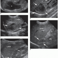


A second sonographic technique used to identify a pneumothorax is with the application of M-mode (motion mode), which is used to detect motion along a select line of interrogation (line of site). In this technique, motion creates waves or curves, and stillness creates straight horizontal lines. A tracing is displayed by placing the M-mode cursor on the pleura between the ribs. The M-mode will reveal parallel lines above the pleural line corresponding to the motionless parietal tissue of the chest wall. In the presence of sliding, a homogeneous granular (sandy) pattern is seen below the pleural line because of the corresponding constant motion of the underlying lung. This normal lung sliding motion has the appearance of a sandy beach intersecting with rolling waves and is known as the “seashore sign” (Fig. 25-6C, D).33,34 In the case of pneumothorax with absent normal sliding, the M-mode reveals a series of parallel horizontal lines, suggesting complete lack of movement both over and under the pleural line. This pattern is known as the “barcode sign” or “stratosphere sign.”33, 34 and 35 Although absent lung sliding suggests pneumothorax, it can occur in the presence of many other conditions, such as main stem intubation, acute respiratory distress syndrome, or pleural adhesions.37

If a pneumothorax is suspected, one should attempt to document the size or extent of the pneumothorax by localizing the point on the chest wall, where the normal lung pattern can be seen. The “lung point” sign determines where the visceral pleura begins to separate from the chest wall at the margin of the pneumothorax. At this point, both absent

and normal lung sliding can be demonstrated between the pneumothorax and the normal lung.27 At the lung point position during expiration, no sliding is seen; but, with inspiration, the lung inflates and the visceral pleura moves up in apposition with the parietal pleura beneath the probe and sliding is again seen.35 The ability to demonstrate the alternating lung sliding and absence of lung sliding within the same field is diagnostic of pneumothorax with a sensitivity of 66% and specificity of 100%.26, 27 and 28


The incidence of a hemothorax after blunt or penetrating chest injury can be noted sonographically as an anechoic or hypoechoic fluid collection localized to the costophrenic angle.36,37 Visualizing an intact diaphragm inferiorly will allow for the certainty that the fluid collection rests within the pleural cavity (Fig. 25-6E). If there is a pleural cavity fluid collection, the lung may sometimes be identified as a triangular structure superior to the diaphragm and should display rhythmic movement corresponding to the patient’s respirations.


Thorax

Lung sonography is effective in deciphering between the differential diagnosis of acute pulmonary edema and acute respiratory failure. Sonographic examinations help practitioners differentiate between pneumonia, hemothorax, lung contusions, and pneumothorax, and better identify the best location for tube thoracostomy or thoracentesis guidance. During the COVID-19 pandemic, lung sonography has proven useful in the diagnosis and management of patients from the point of triage to intensive care.


Biliary Examination


POCUS is a reliable and valuable tool in identifying biliary tree disease in the acute patient.40 Using sonography avoids ionizing radiation, allows for a rapid assessment, and is highly sensitive and specific for gallbladder disease.40 The evaluation of the entire biliary tree allows for the assessment of cholelithiasis, the presence of biliary sludge within the gallbladder lumen, gallbladder wall thickening, pericholecystic fluid, a more accurate sonographic Murphy sign, and/or bile duct dilatation.

The absence or presence of gallbladder stones is often the primary purpose of utilizing POCUS to evaluate the biliary tree for acute cholecystitis. However, acalculous cholecystitis should be considered with the appropriate clinical presentation, including sonographic findings of gallbladder wall thickening, pericholecystic fluid, and a sonographic Murphy sign. The gallbladder should be evaluated in both the longitudinal and transverse planes by sweeping from the neck inferiorly to the fundus superiorly. The gallbladder wall should be measured, and the gallbladder fossa must be examined for fluid collections and edema.


Biliary Examination Preparation

As is the case with most abdominal examinations, a 3.0-to-5.0-MHz frequency curvilinear transducer is used to optimally evaluate the entire organ. Alternatively, a similar frequency phased array probe can be used. Ideally, the patient should be fasting 6 to 8 hours prior to sonographic assessment because typically this avoids the gallbladder from contracting. However, in the POC setting, this may not always be an option because it would make the gallbladder appear contracted and harder to find.

Because the gallbladder is not fixed to body walls like other gastrointestinal organs, it can have a variety of positions in the right upper quadrant, and the patient can be repositioned from a supine into a left lateral decubitus position, to allow for better visualization of the gallbladder by moving it more to the midline. This repositioning can also aid in visualizing mobile stones or sludge as they shift location with patient movement (Fig. 25-7A). If possible, the patient can be asked to hold the right arm above the
head to widen the intercostal spaces. If the ribs are still blocking the view, the patient can be asked to hold a deep breath to further widen the intercostal spaces. Additionally, the probe can be rotated slightly obliquely to align with the intercostal spaces.


Biliary Imaging

To identify the location of the gallbladder, the probe can be positioned on the patient’s right in the anterior-axillary line at the level of the 10th to 11th intercostal spaces or around the mid-epigastric region, with the indicator facing the patient’s head. The gallbladder neck is attached to the main lobar fissure (MLF), thus this structure can be used as a landmark to find the gallbladder. Once the MLF is identified, you can tilt (or fan) the tail of your probe in either direction slowly to find the gallbladder (Fig. 25-7B).

Identifying the common bile duct (CBD) and intrahepatic ducts can be challenging, often requiring significant practice and training in POCUS. As seen in Figure 25-8, when the gallbladder is found, the portal vein would be seen in the leading edge of the screen, often referred to as the “exclamation sign.” The portal triad (CBD, hepatic artery, and portal vein) can be seen in the short axis. This view is also known as the “Mickey Mouse Sign,” with the CBD and hepatic artery, as the “ears,” and the portal vein, as the “head,” making up the structure (Fig. 25-8).

The CBD is located anterior to the main portal vein in the porta hepatis. The inside diameter of the CBD should be measured and assessed for choledocholithiasis or any masses. Color Doppler should be used to decipher the difference between any similarly appearing vascular structure and the CBD. Additional information about the performance of biliary ultrasound, images, and its findings can be found in Chapter 8.


Renal Examination

Patients presenting with undifferentiated flank pain can be rapidly assessed using POCUS with good accuracy and without the exposure to radiation or contrast injection.40, 41 and 42 Focused renal sonography is often utilized to detect and grade hydronephrosis and can be used to assess for renal calculi with or without obstruction of flow.

Only gold members can continue reading. Log In or Register to continue

Stay updated, free articles. Join our Telegram channel

Dec 10, 2022 | Posted by in ULTRASONOGRAPHY | Comments Off on Point-of-Care

Full access? Get Clinical Tree

Get Clinical Tree app for offline access