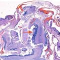
Fig. 19.1
Three-dimensional ultrasound and three-dimensional MRI demonstrated macroglossia (arrow)
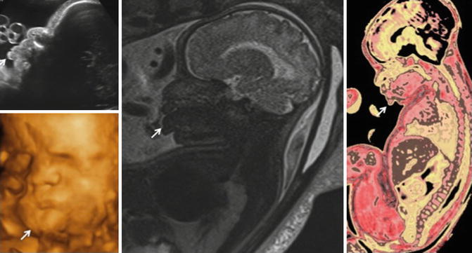
Fig. 19.2
Two-dimensional ultrasound, three-dimensional ultrasound, and fetal MRI demonstrated macroglossia (arrow)
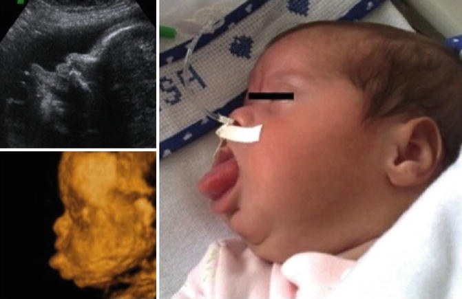
Fig. 19.3
Two-dimensional ultrasound, three-dimensional ultrasound, and postnatal photograph showed macroglossia
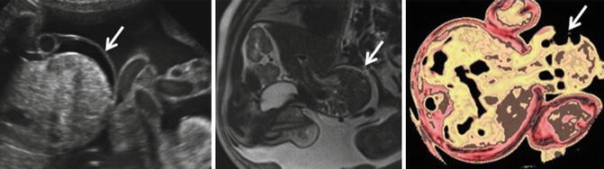
Fig. 19.4
Two-dimensional ultrasound and fetal MRI detected a large (arrow)

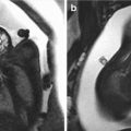
Fig. 19.5
Doppler ultrasound confirmed a left-sided fetal abdominal wall defect and three-dimensional ultrasound clearly rendered the omphalocele (arrows)
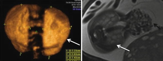
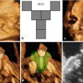
Fig. 19.6
Three-dimensional ultrasound and fetal MRI detected markedly enlarged kidneys (arrows). Note exomphalos on fetal MRI image (black arrow)
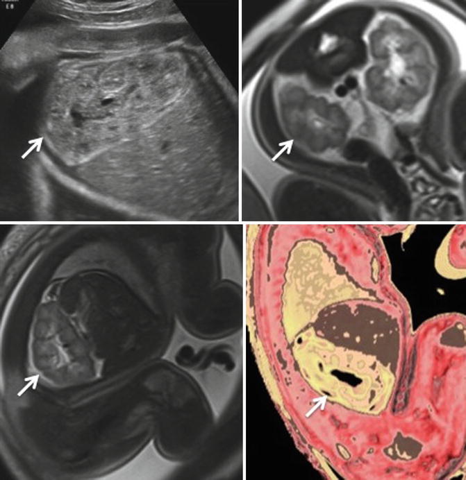
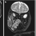
Fig. 19.7
 Congenital Subcutaneous Mixed Venous-Lymphatic Orofacial Malformation Associated with Macroglossia: Prenatal Diagnosis with Ultrasound and Fetal MRI
 The Genetics of Facial Cleft
 Median Cleft Lip and Palate, Cutaneous Nasal Polyps, and Corpus Callosum Lipoma: A Case of Pai Syndrome Associated with Ventricular Septal Defects
 Acromelic Frontonasal Dysplasia (Median Cleft Face Syndrome)
 Magnetic Resonance Imaging (MRI) in the Evaluation of the Fetal Face
 The Role of 2D/3D/4D Ultrasound in the Prenatal Assessment of Cleft Lip and Palate


Two-dimensional ultrasound and fetal MRI detected markedly enlarged kidneys (arrows)
Related posts:
Congenital Subcutaneous Mixed Venous-Lymphatic Orofacial Malformation Associated with Macroglossia: Prenatal Diagnosis with Ultrasound and Fetal MRI
The Genetics of Facial Cleft
Median Cleft Lip and Palate, Cutaneous Nasal Polyps, and Corpus Callosum Lipoma: A Case of Pai Syndrome Associated with Ventricular Septal Defects
Acromelic Frontonasal Dysplasia (Median Cleft Face Syndrome)
Magnetic Resonance Imaging (MRI) in the Evaluation of the Fetal Face
The Role of 2D/3D/4D Ultrasound in the Prenatal Assessment of Cleft Lip and Palate
Stay updated, free articles. Join our Telegram channel
Full access? Get Clinical Tree
