Localization: About half of the cases are localized in the skeleton of the trunk and skull (particularly jaws). The other half occurs in the long bones (metaphysis or diaphysis). Not rarely (around 20 %) involves two or more adjacent or distant bones.
Clinical: Essentially local: pain, swelling, pathological fracture, and neurological deficits in vertebral localization. Not uncommonly symptoms are mild and of long duration.
Imaging: On X-rays: small, diffuse, ill-defined lytic lesions that give a permeative pattern are the most frequent appearance of L. It is difficult to see the end of the lesion. Often a moth-eaten radiographic pattern is observed in L where lytic lesions are greater and spotted. The cortex may be broken or destroyed with a large mass in the soft tissue mimicking a bone sarcoma, and sometimes, the cortex can be thickened mimicking an osteomyelitis. Usually, a periosteal reaction is absent, but in rare cases a specular or lamellar reaction may be observed, and X-rays may mimic those of an osteosarcoma or an Ewing sarcoma. At times, L has an eccentric aggressive lytic lesion like a metastasis, a pure well-defined lytic lesion like a benign bone tumor, or a bubbly, diffused lytic lesions like a myeloma. More rarely, NHL can appear sclerotic, but a sclerotic appearance is more common in H lymphoma, and it may be the first sign of the disease when changes in the lymph nodes are still not very evident. In 30 % of cases, a pathologic fracture is the first appearance of L, and it may occur both in lytic and in sclerotic lesions. On bone scan an uptake shows the lesion, and it defines better than X-rays the real extension of the disease. CT scan shows the typical permeative or moth-eaten radiographic features of L, the mottled appearance due to reactive hyperostosis, and it is very useful to evaluate the relationship with the neurovascular bundles and the involvement of the joint and for the staging of the lesion showing lung metastasis or secondary localization to lymph nodes. MRI is not characteristic in L of long bones. It shows the intramedullary extension of the lesion and the relationship with the joint and the soft tissue, but MRI is really necessary in L of the spine because it can show exactly spinal cord compression and the extension of the disease.
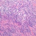
Histopathology: Viable tumor is grayish, soft, and encephaloid. Hemorrhage, necrosis, and colliquation (sometimes with the impression of purulence) are frequent. Tumor manifests a prominent permeation and inflammatory response. Histologically the tumor is composed by sheets of dense roundish cells, clearly separated from each other. Cytoplasm is scant or abundant, pale to clear. Nuclei are large, pleomorphic, often with a vesicular appearance. Chromatin ranges from loose and granular to heavily clumped. Nucleoli are irregular in size, sometimes prominent. Mitoses are usually frequent, sometimes atypical. Reticulin fibers surround small groups of cells and even single cells. PAS does not reveal cytoplasmic glycogen. Immunostains (in undecalcified specimens) are positive for B and T cell markers, immunoglobulins, and common leukocyte antigens. Inflammatory cells are frequently spread in and around the tumor, possibly obscuring the malignant cell population. The majority of primary lymphomas of bone are of intermediate or high histological grade of malignancy. Regarding the phenotype, the majority are B cell lymphomas.
Course and Staging: Often it has a slow course. Pulmonary metastases occur rarely. When lymphoma is suspected or diagnosed, it is necessary to stage the lesion, by studying the skeleton, the regional and distant lymph nodes, the abdominal and thoracic organs and sites, and the central nervous system. Usually NHL remains localized in the bone and it may heal. At times, it may diffuse to regional lymph nodes and it also may heal. In other cases it is present a second or many bone localizations that may involve only regional lymph nodes. Finally, there are cases of systemic NHL that begins from the bone and then diffuse in other sites. At 10 years, the survival is increased from 30 to 60–80 % due to the introduction of polychemotherapy in the treatment protocols of L.
Related posts:


Stay updated, free articles. Join our Telegram channel

Full access? Get Clinical Tree


