67 Proton Spectroscopy (Theory)
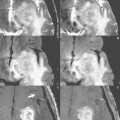
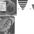
The visual assessment of mass lesions is accomplished in a variety of ways after an MR exam. A few examples of these techniques include the displacement of normal anatomy, the difference in molecular mobility affecting the relaxation times and thus image contrast, and finally the enhancement pattern after contrast administration. In addition to viewing anatomic structure, MR offers the possibility to “visualize” the chemical environment via MR spectroscopy, examining the metabolism of areas in question. Figure 67.1 depicts a lesion (A) examined with a routine imaging sequence, in this instance FLAIR (B), followed by a spectroscopic assessment. The results of the spectroscopy measurement (C,D) demonstrate the elevated level of choline and decreased level of N-acetylaspartate (NAA) typical of malignant lesions. This information can be used to further clarify ambiguous imaging findings.
Stay updated, free articles. Join our Telegram channel

Full access? Get Clinical Tree


