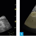
Fig. 1: Information provided by the typical B-mode image.
On the monitor an ultrasound image yields various types of information.
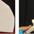
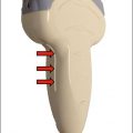
The picture detail (shaded light green here) is shaped like the filter of a coffee percolator. Close to the probe, ultrasound waves are emitted in a fan-shaped manner from the convex surface of the probe to the periphery. The picture detail on the upper left shows a logo, a dot or something similar, which symbolizes the position of the probe marker on the image.
Various technical data are given on the right side of the image, such as “Frq 4.0 MHz” – which means that one is working with an ultrasound frequency of 4 MHz, or “D 14 cm” – which means that the penetration depth of the image is 14 cm. These data are not very significant in the beginning, but as one’s training progresses, they help to focus the device rapidly and handle it in an optimal way.
The scale on the right side of the image is much more important. Resembling a ruler, it expresses the depth of penetration and gives an idea of sizes and dimensions on the monitor. Depending on the depth of penetration, individual structures may appear smaller or larger than one expects them to be. Therefore, correct setting of depth and knowledge of penetration depth on the image are essential aspects of one’s skills.
Picture detail, pictogram showing the sectional plane and penetration depth or the scale constitute essential image data.
Related posts:

Stay updated, free articles. Join our Telegram channel

Full access? Get Clinical Tree


