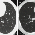
Fig. 21.1
Primary syndrome. CT scanning demonstrates a small quantity of spot-like shadows at the lateral segment of the right middle lobe with fusion of some spot-like shadows, enlarged right hilum that is caused by right hilar lymphadenectasis, and unobstructed adjacent bronchus
Cast Study 2
A female patient aged 20 years was diagnosed with primary pulmonary tuberculosis (primary syndrome).
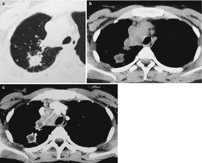
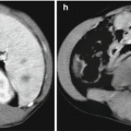
Fig. 21.2
Primary syndrome. (a) CT scanning demonstrates irregular nodular shadows at the apical segment of the right upper lobe, surrounding spot- and cord-like shadows, and flakes of shadows near mediastinum that connect to the mediastinum. (b) CT scanning demonstrates uneven density of the nodules at the right upper lobe, low-density area in the parenchyma, lymphadenectasis at the 2R area of the mediastinum, poorly defined surrounding adipose space, and connection of some adipose spaces with intrapulmonary lesions. (c) Contrast scanning demonstrates marginal enhancement of the nodules at the right upper lung, central low-density area with no enhancement, and uneven enhancement of mediastinal lymph nodes
Intrathoracic Lymphatic TB
Due to rapid absorption of primary lesion in the lung but relatively slow absorption of hilar and mediastinal lymphatic lesion, primary tuberculosis is only demonstrated as enlarged hilar and/or mediastinal lymph nodes, which is known as intrathoracic lymphatic tuberculosis. Hilar lymphatic tuberculosis can be further divided into the following two types: inflammatory lymphatic tuberculosis, with enlarged lymph node and surrounding inflammatory infiltration, and mass lymphatic tuberculosis, with absorption of inflammatory lesion surrounding the lymph node and well-defined boundary of the lymph node.
Hilar Lymphatic TB
Mass Lymphatic Tuberculosis
The lesion of mass type is commonly located at the right hilum, specifically superior margin of the right hilum and the angular area of the right hilum around the right inferior pulmonary artery. The lesion at the left hilar area is commonly located at the superior and exterior margin of the left pulmonary arterial arch and around the left inferior pulmonary artery.
The fully exposed lymph node is nodular, round, or oval in shape with well-defined intact boundary. The partially exposed lymph node is half arch in shape protruding towards the lung field due to its partial overlapping with the hilar vascular vessels. The fusion of large lymph nodes is demonstrated like a mass, with its exterior margin lobulated like plum petals. Plain CT scanning demonstrates some adipose spaces between the lymph nodes, based on which the mass can be distinguished to be formed by fused nodules.
The density of the lesion is homogeneous or heterogeneous (caseous necrosis and liquefaction within lymph lodes). Even calcification can be found.
Contrast CT scanning demonstrates less enlarged lymph nodes with slightly even enhancement and most enlarged lymph nodes with ring-shaped enhancement (peripheral granulation of the lymph node is demonstrated as a ring-shaped or marginal enhancement by contrast scanning, and central necrosis and liquefaction is demonstrated with no enhancement). In the cases with fusion of multiple lymph nodes into large mass, separated enhancement is demonstrated. The enhancement degree of enlarged lymph nodes is markedly distinct from that of blood vessels in the adjacent hilar area (Fig. 21.3).
Inflammatory Lymphatic TB
Inflammatory lymphatic TB shares common radiological demonstrations with mass lymphatic TB.
Due to caseous necrosis and capsular rupture of some lymph nodes, these lymph nodes are demonstrated with rough and poorly defined margin. Otherwise, due to the exudative lesion around the lymph node, the marginal density is demonstrated to be lightened and blurry.
The major vascular branches at the hilar area are mostly demonstrated with smooth blood flow, well-defined and smooth vascular lining, and occasional enlarged lymph nodes. In the cases with the adjacent bronchus involved or with concurrent adjacent bronchial TB, the involved bronchial segment is subject to thickened bronchial wall and narrowed bronchial lumen.
Mediastinal Lymphadenectasis
The lesion is commonly located at the paratracheal area and tracheobronchial region, especially the right lung.
For the cases with well-defined lymph node, pathological examination indicates intact capsule encapsulating the swollen lymph node. X-ray demonstrates the swollen lymph node in a half arch shape protruding towards the lung field, with wavelike mediastinum. Plain CT scanning demonstrates round- or oval-shaped lymph node with surrounding adipose space, especially prevascular space. There are also swollen lymph nodes anterior to the right trachea and posterior to the vena cava, at the left aortic-pulmonary window and in the right azygoesophageal recess. For the cases with unclearly defined lymph node, its surrounding adipose spaces are partially absent and partially present. The pathological examination indicates caseous necrosis of the lymph node and its penetration out of its capsule. For the cases with fusion of multiple lymph nodes into irregular mass, the mass is commonly located between the major vascular vessels in the mediastinum.
By plain CT scanning, the density of most lymph nodes is homogeneous, and the density of rare lymph nodes is uneven due to caseous necrosis and liquefaction. In some cases, the lymph nodes are demonstrated with internal or marginal calcification. Contrast CT scanning demonstrates homogeneous enhancement of the small lymph nodes, peripheral irregular wall enhancement of large lymph nodes, ring-shaped enhancement of the thin wall, and separated enhancement. By pathology, the enhanced area is indicated to be tubercular granulation tissue with abundant blood flow, and the area with no enhancement is indicated to be mostly caseous necrosis. The separated enhancement is pathologically indicated to be the fusion of multiple lymph nodes containing lesions of caseous necrosis (Figs. 21.4 and 21.5).
Case Study 3
A male patient aged 16 years was diagnosed with intrathoracic lymphatic TB.
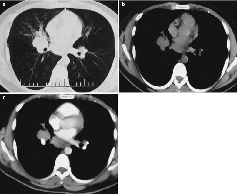
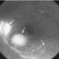
Fig. 21.3
(a, b) Plain CT scanning demonstrates swollen lymph nodes at the right lower lung hilum with nodular shadows and unobstructed right middle bronchus. (c) Contrast CT scanning demonstrates homogeneous enhancement of the hilar major vascular vessels that obviously distinct from its adjacent nodular shadows of lymph nodes. Obstructive pneumonia is demonstrated at the right middle lung lobe
Case Study 4
A male patient aged 21 years was diagnosed with intrathoracic lymphatic TB.
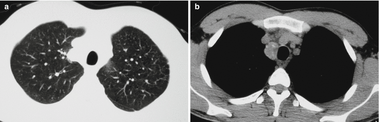
Fig. 21.4
Mediastinal lymphatic TB. (a) CT scanning demonstrates paratracheal nodular shadow at the right upper mediastinum, which is in arch shape protruding towards the lung field. (b) The nodules are in bean shape, with marginal or internal spots of calcification
Case Study 5
A female patient aged 23 years was diagnosed with mediastinal lymphatic TB.
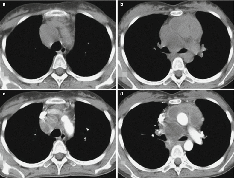
Fig. 21.5
Mediastinal lymphatic TB. (a, b) Plain CT scanning demonstrates swollen lymph nodes at the 3R and 4R areas of the mediastinum, absent adipose space between some of the lymph nodes and vascular vessels, and uneven density. (c, d) Contrast scanning demonstrates heterogeneous enhancement of the lymph nodes, with ring-shaped or separated enhancement
By plain MR imaging, the mediastinal lymph nodes are demonstrated as equal T1 and equal T2 signals, with internal patches of slightly long T1 slightly and long T2 signals. By contrast MR imaging, the mediastinal lymph nodes are demonstrated as nodular, ring-shaped, separated, or uneven enhancement. The demonstrations of no central enhancement, peripheral enhancement, and ring-shaped enhancement are characteristic, with high diagnostic value.
21.7.1.2 Hematogenous Pulmonary TB
Acute Hematogenous Pulmonary TB
It is caused by invasion of a large quantity of MTB into the blood flow for once or several times within a short period of time. It is more common in children and during the stage of primary pulmonary TB.
X-Ray
X-ray demonstrates extensively and evenly distributed miliary sized nodular shadows with increased density. It is characterized by even distribution, even size, and even density, which is known as the three-evens sign. Due to a large quantity of lesions with dense distribution, both lungs are demonstrated with ground-glass opacity. The miliary shadows have a diameter of 1–2 mm and a round or oval shape, with well-defined boundary. In the cases with exudative lesions, the miliary shadows are demonstrated with poorly defined boundary. At the early stage, X-ray only demonstrates enhanced lung markings. After about 2 weeks, typical miliary nodules can be demonstrated. In the advanced stage, the miliary increased density shadows tend to fuse together.
CT Scanning
Pulmonary interstitial miliary nodules are demonstrated with diffusely distributed military shadows at the interstitium of both lungs. The nodules have a diameter of 1–3 mm with even distribution and even density. Most nodules are well defined and rarely with poorly defined boundary. In the cases with no immediate treatment, the nodules may enlarge to a diameter of about 5 mm. The nodules are morphologically irregular and may fuse into focalized consolidation lesion.
Miliary nodules may be complicated by confined ground-glass opacity, with light density and blurry boundary.
Interlobular septal thickening and intralobular reticular shadows are caused by congestion and edema of alveolar septa during the acute stage, which often coexists with ground-glass opacity. Most patients experience their absence after treatment. In some rare patients, they develop into irreversible reticular fibrosis.
Clustering thin wall cyst shadows can be demonstrated at the progressive stage of the conditions in rare patients, which are reversible.
In the patients with acute hematogenous pulmonary TB but receiving delayed or inappropriate therapy, the conditions progress. The caseous substances formed by TB lesions may involve the alveolar cavity and disseminate along the bronchus. The nodular shadows in both lungs can be demonstrated with random and confined distribution of centrilobular branch shadow and the tree bud sign that may be well defined or poorly defined (Figs. 21.6 and 21.7).
Case Study 6
A female patient aged 14 years was diagnosed with acute hematogenous pulmonary TB.
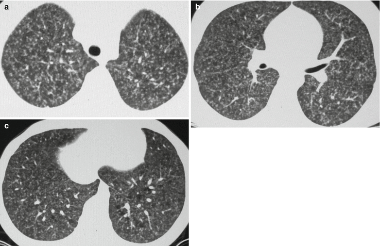
Fig. 21.6
Acute hematogenous pulmonary TB. (a–c) CT scanning demonstrates diffuse miliary shadows at both lungs, with even size, even density and even distribution
Case Study 7
A female patient aged 25 years was diagnosed with acute miliary pulmonary TB. During the recent 10 days, the patients experience fever and cough after abortion, with difficulty expectorating. The fever occurs commonly after noons, with a body temperature fluctuating around 37.8 °C, and the cough is paroxysmal.
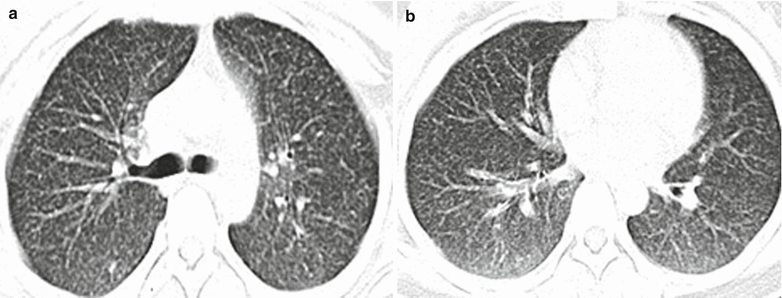
Fig. 21.7
Acute miliary pulmonary TB. (a, b) CT scanning demonstrates diffuse miliary nodular shadows at both lungs with even size, even density, and even distribution
Subacute and Chronic Hematogenous Pulmonary TB
Due to repeated invasions of a small quantity of MTB into the blood flow during a long period of time, subacute or chronic hematogenous pulmonary TB occurs. The source of their dissemination is commonly infected veins after urogenital TB or osteoarticular TB.
X-Ray
X-ray demonstrates three-unevens sign. The size of lesions ranges from miliary to 1 cm in diameter. The density of exudative or proliferative lesions is light, while the density of calcification is dense, with blurry or sharp boundary. The lesions are unevenly distributed at the upper and middle lung fields. The old hard nodular calcifications are mostly distributed at the lung apex and subclavicular area, while the new exudative and proliferative lesions are mostly distributed at the lower lungs.
CT Scanning
CT scanning demonstrates unevenly distributed lesions that are more commonly distributed at the middle and upper lungs, the lesions with different sizes due to fusion of miliary nodules, and the lesions with uneven density due to inner calcification. These findings are known as the three-unevens sign. Compensatory emphysema can be demonstrated between lesions or at the lower lungs (Fig. 21.8).
Case Study 8
A male patient aged 24 years was diagnosed with subacute hematogenous pulmonary TB.
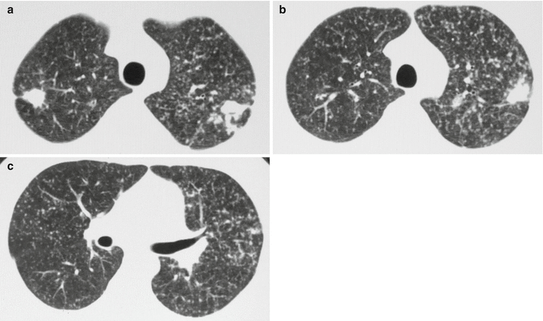
Fig. 21.8
Subacute hematogenous pulmonary TB. (a–c) CT scanning demonstrates diffuse miliary nodules at both lungs, with different size and density. Some miliary nodules fuse together at the upper lobe of both lungs
Secondary Pulmonary TB
Secondary pulmonary TB is a major type of pulmonary TB, and it is the most common type of pulmonary TB.
Infiltrative Pulmonary TB
CT Scanning
CT scanning sometimes demonstrates traces of primary infection of tuberculosis. The calcification in the intrapulmonary primary lesions is demonstrated in spots of and nodular lesions with high density. Peripheral inflammation can be demonstrated around the old lesions, with central high density and peripheral patches and small flakes of light blurry shadows. Hilar and mediastinal lymph nodes are demonstrated with calcification.
The lesions of secondary infection of MTB are commonly located at the apical posterior segment of the upper lung lobe and dorsal segment of the lower lung lobe. Multiple lesions can be found within one pulmonary segment or lobe or within unilateral or bilateral lung fields. The exudative lesions are demonstrated as cloud-like shadow with patches, small flakes, or flakes of shadows with increased density. The density of lesions is even or uneven with blurry boundary. The patches of shadows may fuse into flakes of shadows. The proliferative lesions are demonstrated as spots, small nodules, and patches of shadows with increased density and well-defined boundary. The proliferative lesions rarely fuse together. Caseous necrosis can be found at the lesions to induce low-density or semitransparent cavity, which is demonstrated as a transparent area with different sizes and shapes. Infiltrative TB at different stages can be demonstrated with no wall cavity, thin wall cavity, caseous thick wall cavity, tension cavity, and purified cavity. Around the cavity, spots of small nodular shadows are demonstrated, which are technically known as satellite lesions. Bronchial lesions are commonly found at the unilateral or bilateral middle and lower lung fields disseminated from cavity. The calcifications are demonstrated as high-density spots of linear or patches of shadows with well-defined boundary but in regular or irregular shape. At the middle and advanced stages of infiltrative TB, calcification is commonly demonstrated in the lesions. The finding of calcifications in the lesion or at the margin of lesion greatly facilitates the qualitative diagnosis. Meanwhile the finding of calcification indicates that the conditions are tending to stabilize or be improved. The fibrous lesions are demonstrated as cords like or stellate-like shadows with high density and rough boundary. Within a short period of time, the progress and absorption of the lesions are slower than those of lobar pneumonia, lobular pneumonia, mycoplasma pneumonia, and common viral pneumonia. Accompanying pleural effusion occurs. In the cases complicated by hematogenous disseminated pulmonary TB, in addition to intrapulmonary infiltrative lesions, there are also demonstrations of acute or subacute and chronic hematogenous disseminated pulmonary TB (Figs. 21.9, 21.10, 21.11, and 21.12).
Infiltrative pulmonary TB is demonstrated with common concurrence of at least two types of lesions among exudates, proliferation, caseous necrosis, cavity, calcification, and fibrosis. Radiologically, it is characterized by multiple lesions, multiple densities, and multiple shapes. The typical radiological sign of infiltrative pulmonary TB is lesions at the apical posterior segment of both upper lung lobes.
Case Study 9
A male patient aged 20 years was diagnosed with secondary pulmonary TB (infiltrative pulmonary TB).
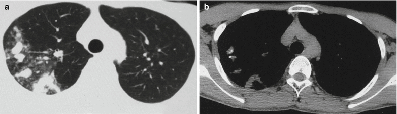
Fig. 21.9
Infiltrative pulmonary TB. (a, b) CT scanning demonstrates spots of small nodular and small patches of shadows at the right upper lung lobe and calcification in the lesions at the apical segment of the right upper lung lobe
Case Study 10
A male patient aged 25 years was diagnosed with secondary pulmonary TB (infiltrative pulmonary TB).
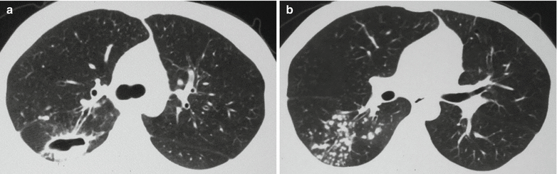
Fig. 21.10
Infiltrative pulmonary TB. (a, b) CT scanning demonstrates a transparent area at the posterior segment of the right upper lung lobe, double rail-like draining bronchi between the transparent area and the hilum, spot-like satellite lesions around the transparent area, and spots of bronchial lesions at the right lower lung lobe
Case Study 11
A female patient aged 77 years complained of cough for over 4 months, with rare phlegm, fatigue, chest distress, and shortness of breath. By sputum smear, acid-fast bacillus is positive.
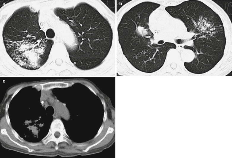
Fig. 21.11
Infiltrative pulmonary TB. (a–c) CT scanning demonstrates flakes and nodular blurry shadows at both lungs, dilation of local bronchial lumen, thickening of local bronchial wall, and calcification of some lymph nodes
Case Study 12
A female patient aged 43 years. By sputum smear, acid-fast bacillus is positive.
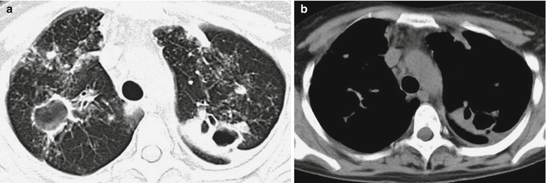
Fig. 21.12
Infiltrative pulmonary TB and pleural effusion. (a, b). CT scanning demonstrates multiple thick wall cavities in different sizes at both upper lung lobes, uneven thickness of the thick wall cavities, multiple small patches and cotton wool like shadows around the lesions, observable tree bud sign, and scattering multiple nodular or cord-like shadows in both lungs. There are also enlarged mediastinal lymph nodes and bilateral pleural effusion in small quantities
MR Imaging
T2WI demonstrates exudative and caseous lesions with high signal, proliferative lesions with moderate signal, fibrosis lesions with low signal, calcification lesions with lower signal, and gas in the cavity with extremely low signal.
Tubercular Lobar Pneumonia and Caseous Pneumonia
Tubercular Lobar Pneumonia
Tubercular lobar pneumonia is an uncommon type of secondary pulmonary TB, which is also known as bronchial pneumonia TB.
By CT scanning, the lesions are commonly located at the upper lung lobe, especially the apical posterior segment. The lesions are commonly demonstrated as large flakes of shadows occupying a whole segment, lobe, or even a lung field. The central area is demonstrated with a large area of consolidation, with light and poorly defined boundary. These demonstrations resemble to those of lobar pneumonia. The large flakes of consolidation are demonstrated with inner air bronchus sign. In some cases, the air bronchus is demonstrated with column-like or bead string-like dilation. The cloud-like shadows are demonstrated with spots of and small nodular high-density lesions. The small nodular shadows are demonstrated at the margin of flakes of shadows, with some marginal lesions being blurry and some others well defined. By radiological examinations, a large flake of lesions is surrounded by many small lesions, which scatter unevenly. Such findings facilitate its differential diagnosis from lobar pneumonia. In the large flake of consolidation or at the marginal flake of consolidation, there are slightly low-density semitransparent areas or small transparent areas, indicating the occurrence of small caseous necrosis and dissolution. The disease progresses rapidly, with infiltration of the lesions, occurrence of surrounding inflammation, and fusion of the lesions. At the affected lung field or contralateral lung field, scattering polymorphic spots of small nodular or patches of TB lesions greatly facilitate its diagnosis (Fig. 21.13).
Case Study 13
A male patient aged 36 years was diagnosed with secondary pulmonary TB (tubercular lobar pneumonia).
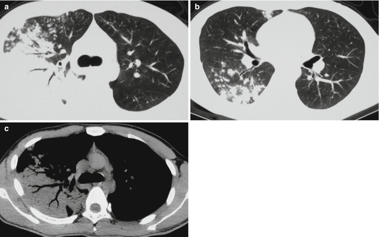
Fig. 21.13
Tubercular lobar pneumonia. (a–c) CT scanning demonstrates a large flake of consolidation at the right upper lung lobe with inner air bronchus sign. The margin of lesions is demonstrated to be poorly defined with small patches and spots of shadows. The middle and lower lobes of right lung are demonstrated with small patches and spots of shadows
Caseous Pneumonia
Caseous pneumonia is the acutest and severest type of secondary pulmonary TB. Some cases of caseous pneumonia are developed from caseous necrosis of exudative lesions due to tubercular lobar pneumonia.
1.
X-ray and CT scanning
Caseous lobar pneumonia is demonstrated with large flakes of cloud-like shadows, which gradually develop into a large flake of consolidation shadow with moderate density. The lesion is commonly located at the upper lung lobe, occupying a whole segment, lobe, or even lung field, with poorly defined boundary. The lesion has uneven density, with extensive multiple worm-bitten-like low-density semitransparent area or transparent area in different sizes at the large flake of consolidation shadow. The cavities are morphologically varied, with irregular, unsmooth, and poorly defined cavity wall. Such a sign is more clearly demonstrated by HRCT. Without timely and effective treatment to control its development, the conditions continue to deteriorate with further expansion of multiple worm-bitten-like cavities in different sizes and their fusion. Consequently, large area of the lung is destructed by formation of huge cavity (Fig. 21.14). Bronchial lesions caused by dissemination can be demonstrated at the homolateral or contralateral lung field. Caseous lobular pneumonia is demonstrated with multiple scattering small flakes, small nodules, and spots of shadows at the unilateral or bilateral upper and middle parts of the lung. The small flakes of shadows may fuse into a large flake. In the flakes of, small flakes of and nodular lesions, irregular lesions of caseous necrosis and dissolution can be found in different sizes to show low-density semitransparent or transparent area. Such a sign is of great significance for the diagnosis. At the middle and advanced stages, the lung tissue is subject to extensive and severe damage to cause shrinkage of the lung lobe and thickening of the adjacent pleura.
2.
MR imaging
The demonstrations of secondary pulmonary TB based on caseous lesions by MR imaging morphologically resemble to those by CT scanning. The lesions of caseous necrosis are demonstrated with moderate or slightly low T1WI signal and heterogeneous high T2WI signal. In the cases with peripheral fiber or granulation tissue, enhancement can be demonstrated, while the caseous necrotic tissue is demonstrated with no enhancement. In the cases with accompanying large calcification, no signaling shadow can be demonstrated.
Case Study 14
A female patient aged 30 years was diagnosed with secondary pulmonary TB (caseous pneumonia).
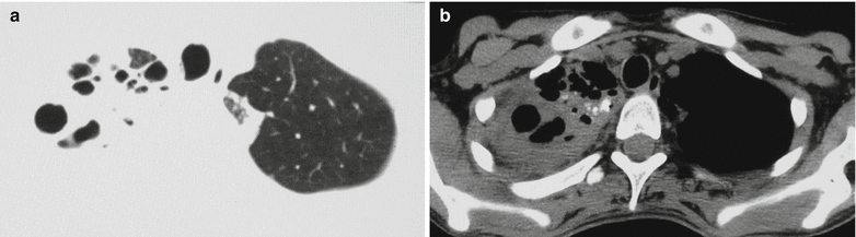
Fig. 21.14
Caseous pneumonia. (a) CT scanning demonstrates large flakes of shadows at the right upper lung lobe with multiple irregular transparent areas in different sizes in the lesions and cord-like shadows at the left upper lung lobe. (b) Spots of calcification are demonstrated in the lesion
Pulmonary Tuberculoma
Tuberculoma refers to the spherical lesion formed by a caseous lesion of pulmonary TB encapsulated by fibrous tissues.
1.
The lesions are commonly located at the apical posterior segment of the upper lung lobe and dorsal segment of the lower lung lobe.
2.
The lesion is mostly singular, but in rare cases, the lesions are multiple.
3.
The lesions are round or oval in shape, those with a diameter being less than 2 cm are referred to as fibrous caseous lesion, and those with a diameter being larger than 2 cm are referred to as tuberculoma.
4.
The contour of lesion is clear and regular, rarely with incisura and lobulation.
5.
The density of the lesion may be homogeneous or with inner semitransparent area due to dissolution. The dissolution area is commonly located near the hilum in the spheric shadow. Cavity can also be demonstrated.
6.
At the lung field near the spherical shadow, scattering proliferative or fibrous lesions are commonly found in spots of small nodular and cord-like shadows, namely, the satellite lesions.
7.
Between the tuberculoma and hilum of the lung, cord-like shadows are demonstrated, which are residual draining bronchi after formation of the tuberculoma derived from the cavity.
8.
Calcification can be found within or at the margin of tuberculoma. The typical demonstration is laminar ring-shaped calcification shadow or scattering spots of calcification.
Case Study 15
A male patient aged 39 years was diagnosed with secondary pulmonary TB (pulmonary tuberculoma).
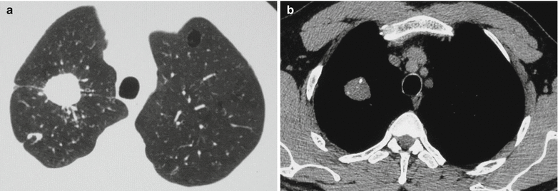
Fig. 21.15
Pulmonary tuberculoma. (a) CT scanning demonstrates round-like nodular shadow at the apical segment of the right upper lung lobe, with a well-defined and smooth boundary, multiple linear shadows in different lengths radiating from the nodule outwards, and spots of satellite lesion around the lesion. (b) Spots of calcifications can be demonstrated in the lesion
Case Study 16
A female patient aged 43 years experienced cough and hemoptysis for several months. By sputum smear, acid-fast bacillus is positive.

Fig. 21.16
Pulmonary tuberculoma, infiltrative pulmonary TB. (a, b) CT scanning demonstrates spherical lesion at the right middle lung lobe, with marginal irregular calcification and well-defined boundary. The lesion is demonstrated with surrounding satellite lesions. And multiple nodules are demonstrated at the right lower lung
Case Study 17
A male patient aged 35 years experienced cough for over 20 days, with no known causes. He coughed up rare sputum that was white and foamy, with no blood and odor. By sputum smear, acid-fast bacillus is positive.
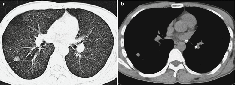
Fig. 21.17
Pulmonary tuberculoma and acute hematogenous pulmonary TB. (a, b) CT scanning demonstrates spherical lesion at the right lower lung lobe near pleura with poor defined boundary and surrounding satellite lesions. There are diffuse miliary TB lesions at both lungs and calcified lymph nodes at the mediastinum and left hilum
Chronic Fibro-Cavity Pulmonary TB
Tuberculosis has a long illness course, with alternated deterioration and alleviation. Local or most of the lung tissue is subject to severe damage, with concurrent extensive proliferation and repair of fiber tissue around the lesion, namely, the occurrence of fibro-cavity.
1.
Irregular fibro-cavities are located at the apical posterior segment of the upper lung lobe or the dorsal segment of the lower lung lobe, namely, the supraclavicular or subclavicular region. Extensive cord-like shadows are demonstrated to surround the lesion. Local lung volume shrinks, commonly leading to elevated hilum at the affected lung. The lung markings are demonstrated like willow. And the trachea and mediastinum are demonstrated to shift to the affected lung.
2.
Both old and new tuberculosis lesions are commonly found at the unilateral upper and middle lung fields, namely, concurrence of various lesions including exudative lesions, proliferative lesions, caseous lesions, cavities, fibrous lesions, and calcification.
3.


Bronchial lesions due to dissemination are commonly found at the middle and lower lung fields of the affected lung as well as at the contralateral lung field.
Related posts:

Stay updated, free articles. Join our Telegram channel
Full access? Get Clinical Tree
