Radiation Cataractogenesis
▪ CATARACTS OF THE OCULAR LENS
The word cataract is used to describe any detectable change in the normally transparent lens of the eye. The effect may vary from tiny flecks in the lens to complete opacification, resulting in total blindness. Cataracts are usually associated with old age or, less commonly, with some abnormal metabolic disorder, chronic ocular infection, or trauma. It is also well known that sufficient exposure to ionizing radiations (such as x- or γ-rays, charged particles, or neutrons) may cause a cataract.
The ocular lens is enclosed in a capsule (Fig. 13.1); the lens itself consists largely of fiber cells and is covered with an epithelium anteriorly. The lens has no blood supply. Dividing cells are limited to the preequatorial region of the epithelium. The progeny of these mitotic cells differentiate into lens fibers and accrete at the equator.
Cell division in the lens continues throughout life, but it is a most curious cellular system in that there is no mechanism for cell removal. If dividing cells are injured by radiation, the resulting abnormal fibers are not removed from the lens but migrate toward the posterior pole; because they are not translucent, they constitute the beginning of a cataract.
▪ LENS OPACIFICATION IN EXPERIMENTAL ANIMALS
Some species of animals, especially the mouse, are very sensitive to radiation as far as lens opacification is concerned. A large proportion of a mouse population naturally develop opacifications as they become older. A dose of a few tens of milligray (mGy) of x-rays or a fraction of 1 mGy of fast neutrons produces readily discernible changes in the lens. As the dose is increased, the latent period (i.e., the time that elapses before an opacity of given severity is evident) becomes shorter. Put another way, radiation advances in time, a process that occurs normally late in life.
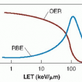
Neutrons and other densely ionizing radiations are very effective at inducing cataracts, as evidenced by several physicists and engineers who developed cataracts as a result of working around high-energy accelerators in the early days before safety procedures were introduced. The relative biologic effectiveness (RBE) of fast neutrons is a strong function of dose, with a value of about 10 pertaining to high-dose levels on the order of several grays relative to x-rays, but rising to 50 or more for small doses of less than 10 mGy. Worgul and his associates have reported similar RBEs for lens damage in rat eyes exposed to accelerated heavy ions. The increase in RBE at low doses is caused largely by the sharply declining effectiveness of x-rays with decreasing dose, rather than an increase in effect per unit dose of neutrons or charged particles.
▪ CATARACTS IN HUMANS
Radiologists have known for many years that the lens of the eye may be damaged by radiation. A study of patients treated with x- or γ-rays in which a proportion of the dose reached the eye has provided some insight into radiation cataractogenesis in humans. Figure 13.2 shows a typical cataract in a patient who had undergone radiotherapy. An early radiation cataract viewed through an ophthalmoscope may appear
as a dot, usually situated at the posterior pole. As it enlarges, small granules and vacuoles appear around it. With further enlargement, to the point at which the opacity is several millimeters in diameter, it may develop with a relatively clear center, so that it is shaped like a doughnut. At the same time, granular opacities and vacuoles may appear in the anterior subcapsular region, usually in the pupillary area. This sequence of events is not unique to radiation, but its appearance in a person with a radiation history strongly suggests radiation as the causative agent. Similarly, an absence of this sequence of events would exclude radiation as a cause. In other words, although it is never possible to state unequivocally that a given cataract is radiation induced, it is possible to say with some certainty that some cataracts—for example, nuclear cataracts—do not have a radiation etiology. Depending on the dose, the cataract frequently remains stationary at this stage, confined to the posterior subcapsular region. If it continues to progress, it becomes nonspecific and cannot be distinguished from other types of cataracts.
as a dot, usually situated at the posterior pole. As it enlarges, small granules and vacuoles appear around it. With further enlargement, to the point at which the opacity is several millimeters in diameter, it may develop with a relatively clear center, so that it is shaped like a doughnut. At the same time, granular opacities and vacuoles may appear in the anterior subcapsular region, usually in the pupillary area. This sequence of events is not unique to radiation, but its appearance in a person with a radiation history strongly suggests radiation as the causative agent. Similarly, an absence of this sequence of events would exclude radiation as a cause. In other words, although it is never possible to state unequivocally that a given cataract is radiation induced, it is possible to say with some certainty that some cataracts—for example, nuclear cataracts—do not have a radiation etiology. Depending on the dose, the cataract frequently remains stationary at this stage, confined to the posterior subcapsular region. If it continues to progress, it becomes nonspecific and cannot be distinguished from other types of cataracts.
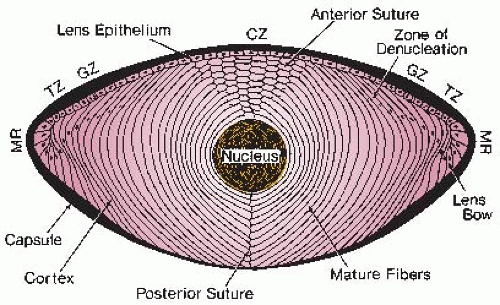 FIGURE 13.1 Diagram of a sagittal section of a human lens, illustrating the various cellular relationships. Cells are produced by mitosis in the germination zone (GZ) of the epithelium. They begin to differentiate into lens fibers at the meridional rows (MR) and accumulate at the equator. Cells in the central zone (CZ) do not normally divide. (Adapted from Merriam GR, Worgul BV. Experimental radiation cataract: its clinical relevance. Bull NY Acad Med. 1983;59:372-392, with permission.) |
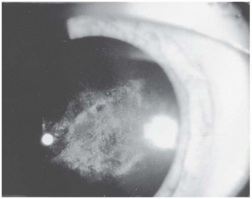 FIGURE 13.2 Cataract in the posterior subcapsular region 4 years after a dose of 24 Gy of x-rays to a patient on radiotherapy. (From Merriam GR, Worgul BV. Experimental radiation cataract: its clinical relevance. Bull NY Acad Med. 1983;59:372-392, with permission.) |
▪ THE DEGREE OF OPACITY
Related posts:

Stay updated, free articles. Join our Telegram channel

Full access? Get Clinical Tree


