1 Radiographic Imaging Techniques and Interpretation
For the evaluation of sinonasal pathology, conventional radiography (plain films) is still a fast and frequently used tool. However, for more accurate evaluation, computed tomography (CT) and magnetic resonance imaging (MRI) are the modalities of choice. These modalities provide much more information about the localization of pathology, its relation to adjacent structures, and expansive or infiltrative characteristics—factors that play an important role in therapeutic decisions and in planning surgery.
With CT, the bony structures can be well evaluated, especially when evaluating bony outlines. Different settings focusing on bone or soft tissues, as well as the use of contrast agents, can refine the differential diagnosis. Using MRI and its different sequences, characteristics of soft tissues as well as any extension into adjacent structures can be well assessed. The relevant differences between CT and MRI are summarized in Table 1.1. For a more extended discussion of the physical characteristics of CT and MRI, as well as their differences, the reader is referred to other manuals listed at the end of this book.
Differentiating Characteristics on CT and MRI
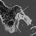
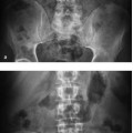
For a detailed evaluation of fine structures on CT, slice thickness and CT settings, as well as the use of intravenous contrast, are important Figure 1.1 shows an intraorbitally located subperiosteal abscess resulting from ethmoiditis. The intraorbital abscess may well be missed in the bone setting of Fig. 1.1a, since differences between the soft tissues are displayed poorly with contrast, even with the use of intravenous contrast as in this image.
As well as the parameters mentioned for the evaluation of CT, such as slice thickness and use of contrast, tissue differentiation on MRI can be refined by the choice of pulse sequence. In general, interpretation of the most frequently used modality, the turbo spin-echo shown in Table 1.2, is of practical value. For a more specific classification and other modalities of MRI, the reader is referred to other manuals or their own department protocol.
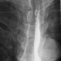
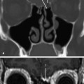
Figure 1.2 a,b illustrates the complementary tissue differentiation by the two modalities of the same tissue slice in a patient with a T4N0 sinonasal carcinoma.
Table 1.1 Practical differences between CT and MRI
| Computed tomography | Magnetic resonance imaging | |
|---|---|---|
| Utilizes radiation with its inherent danger of oncogenic potency, and potential to damage the eye lens and thyroid gland | Does not require radiation, but there is sometimes a slight local rise of tissue temperature | |
| Fast, widely available | Time-consuming, less widely available | |
| Axial slices, with the possibility of coronal and sagittal reconstructions | Original slices in all directions |