2 Radiologic Anatomy of the Temporal Bone
Today, computed tomography (CT) is the most frequently used tool to evaluate the temporal bone. It is used not only to detect pathology, but also as an evaluation tool preoperatively and during operative procedures. Preoperatively, CT may be helpful in decisions about the most optimal surgical approach and may help minimize complications during surgery.
Magnetic resonance imaging (MRI) is used for detecting retrocochlear pathology and intracranial pathology, although it may have a complementary value in evaluating the patency and fluid contents of the inner ear structures. Radiologic anatomy with regard to MRI will be discussed in Chapter 4.
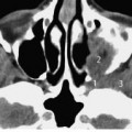
Plain films of the skull to evaluate the temporal bone consist of projections described by Schüller and Stenvers. These projections have been used as a screening tool, but are no longer considered very useful. The Stenvers projection is useful after cochlear implantation to evaluate the position of the electrode in case of difficult insertion (see Fig.2.1). In trauma cases, plain films give an impression of the integrity of the system, but this is usually evaluated with CT.
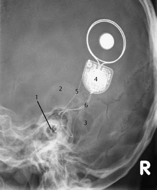
Fig.2.1 Traditional Stenvers radiographic projection. This projection is currently used for, e.g., cochlear implantation to demonstrate the position of the electrode (1). In addition, the radiograph shows the cartilage of the outer ear (2), degree of pneumatization and aeration of the mastoid (3), the processor of the implant (4), the reference electrode (5), and the active electrode (6).
Evaluating a CT Scan of the Temporal Bone
For a systematic and complete analysis, a CT scan of the temporal bone should be evaluated in a standardized manner. An axial evaluation is best started by using cranial slices consisting of only a few structures that can be recognized more easily, to more caudal slices with a somewhat more complex anatomy. Coronal evaluation is best started anteriorly with recognition of the mandibular structures, followed by evaluation of the more posterior slices.
A comprehensive list of evaluation points is given below. In practice, documentation of the findings must be accurate, mentioning not only pathological findings but also the state of essential normal structures. Of course, not every point in the list below needs to be noted in the medical file.
Mastoid Region
• Presence and extension of the pneumatization.
• Contents of the mastoid cells (air, fluid, soft tissue).
• Position of sigmoid sinus.
• Appearance of the bony trabecular structures: intact, destruction.
• Aspect of the lateral bony border of the mastoid.
• Aspect of the bony borders of the posterior and middle fossa.
• Position and integrity of the vertical part of facial nerve canal.
Middle Ear
• Contents: loss of aeration.
• Localization and aspect of masses.
• Ossicular chain: dislocation, destruction, ankylosis.
• Appearance of the round and oval window niche.
• Aspect of the horizontal part of the facial nerve canal.
• Aspect of the region of the geniculate ganglion.
• Aspect of the ear drum: thickened, retracted.
• Position and aspect of the carotid artery, bony coverage.
• Position and aspect of the jugular bulb, bony coverage.
Inner Ear and the Petrous Apex
• Cochlea: demineralization of bony capsule, presence of the modiolus, aspect of cochlear windings and the cochlear lumen, ossification.
• Appearance of vestibular parts and semicircular canals, intact borders, ossification.
•
Related posts:
Stay updated, free articles. Join our Telegram channel

Full access? Get Clinical Tree


