, Valery Kornienko2 and Igor Pronin2
(1)
N.N. Blockhin Russian Cancer Research Center, Moscow, Russia
(2)
N.N. Burdenko National Scientific and Practical Center for Neurosurgery, Moscow, Russia
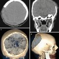
During the “pre-CT period,” the diagnosis of brain lesions was based on the analysis of clinical symptoms, X-ray images, EEG data, radionuclide studies, cerebral angiography, air encephalography, ventriculography, and lumbar puncture (Moniz 1934; Engeset and Kristiansen 1940; Shlifer 1941; Lima 1950; Ecker and Riemenschneider 1955; Decker 1960; Robertson 1967). Despite such a large arsenal of diagnostic methods, diagnostic errors occurred quite a lot. Thus, according to Nisce et al. (1971), autopsies did not detect any brain tumors in 48 of 136 patients who underwent radiation therapy for brain metastases diagnosed based on the conclusions of radiological diagnostic methods. Of course, it is impossible to detect a tumor in the brain parenchyma and estimate its volume, structure, as well as the number of lesions based on X-ray findings. However, metastases in the bone structures of the skull can be diagnosed by X-ray imaging. Thus, metastases of malignant tumors of various origins in the cranial vault can be comparatively easily found on X-ray in frontal and lateral views. Figure 6.1 shows bone lesions of the cranial vault in breast cancer metastases. Metastatic changes in the cranial bones sometimes require X-ray images in special views in addition to the standard ones.


