4 Radiologic Anatomy of the Skull Base
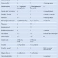
The skull base can be evaluated by computed tomography (CT), which will demonstrate the bony structures of the skull base with its foramina and fissures for vessels and cranial nerves, the temporal bone, and sinonasal cavities. Magnetic resonance imaging (MRI) will demonstrate the contents of the foramina and fissures as well as the intracranial soft tissues. CT or MRI may provide enough information individually to demonstrate and classify the pathology in this area, however, when used together these modalities can be complementary and define even better the invasion and destruction of (bony) structures of the skull base by soft-tissue masses.
Radiologic Evaluation Points of the Skull Base
Computed Tomography
• Bony outline of the outer skull.
• Bony outline of the intracranial skull base.
• Temporal bone structures: internal auditory canal, vestibular and cochlear aqueduct, apex.
• Foramina: ovale, spinosum, jugular, rotundum.
• Large vessels: carotid artery, sigmoid sinus, jugular bulb.
• Supraorbital fissure and orbital structures.
• Infratemporal fossa, sphenoidal bone, clivus.
• Clinoid processes, sella and pituitary fossa.
• Features of the pathology: expanding or invasive growth pattern.
Magnetic Resonance Imaging
• Intracranial brain structures: cerebrum, cerebellum, pons and brainstem, ventricles, dural outlines.
• Vascular structures: transverse sinus, sigmoid sinus and jugular bulb, superior petrosal sinus, carotid artery, vertebrobasilar system, anterior and posterior inferior cerebellar arteries (AICA and PICA).
• Temporal bone: fluid contents (T2-weighted MR image) of inner ear structures and internal auditory canal, appearance of the cochlear, vestibular (inferior and superior), and facial nerves.
• Intensities on T1-weighted and T2-weighted MR images, with contrast, and possible asymmetry.
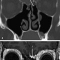
Evaluation of the Skull Base on Axial CT Slices in a Craniocaudal Sequence
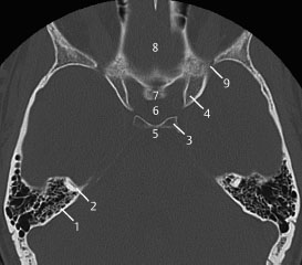
Fig.4.1 CT slice.
1 Temporal bone
2 Anterior semicircular canal
3 Posterior clinoid
4 Anterior clinoid
5 Dorsum sellae
6 Pituitary fossa
7 Tuberculum sellae
8 Fovea ethmoidalis (cranial nerve I in anterior cranial fossa)
9 Superior orbital fissure
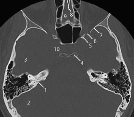
Fig.4.2 Axial CT slice.
1 Subarcuate canal and artery
2 Posterior cranial fossa
3 Middle cranial fossa
4 Posterior clinoid process
5 Anterior clinoid process
6 Superior orbital fissure (cranial nerves III, IV, VI, and a part of V)
7 Sphenoid bone
8 Crista galli
9 Fovea ethmoidalis
10 Region of the cavernous sinus and internal carotid artery
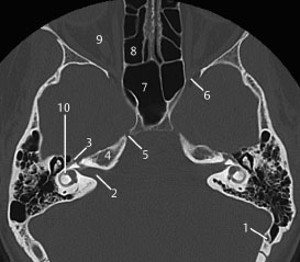
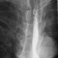
Fig.4.3 Axial CT slice.
1 Emissary vein
2 Internal auditory canal (cranial nerves VII and VIII)
3 Geniculate ganglion
4 Petrous apex
5 Foramen for the ophthalmic nerve (part of the trigeminal nerve)
6 Superior orbital fissure
7 Sphenoid sinus
8 Ethmoid sinus
9 Optic nerve (cranial nerve II)
10 Horizontal semicircular canal and vestibule
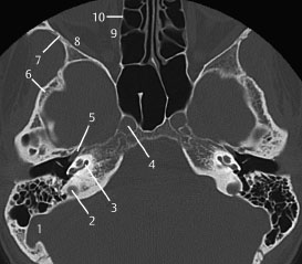
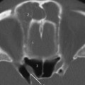
Fig.4.4 Axial CT slice.
1 Sigmoid sinus
2 Roof of the jugular bulb
3 Cochlea (basal turn)
4 Internal carotid artery
5 Eustachian tube
6 Temporal bone (squamous part)
7 Greater wing of sphenoid
8 Lateral rectus
9 Medial rectus
10 Lamina papyracea

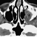
Fig.4.5 Axial CT slice.
1 Jugular bulb
2 Cochlear aqueduct
3 Basal turn of cochlea
4 Internal carotid artery
Related posts:
Stay updated, free articles. Join our Telegram channel

Full access? Get Clinical Tree


