- •
Contrast-enhanced renal MRA is an accurate, noninvasive technique for detecting renal artery stenosis.
- •
Phase contrast MRA techniques can be used to assess the hemodynamic significance of a stenosis.
- •
MRA plays an important role in the evaluation of vascular complications following renal transplant.
- •
Non-contrast-enhanced MRA techniques can be used to noninvasively evaluate the renal vasculature in patients who cannot receive MR contrast agents.
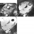
A 65-year-old woman was referred for renal magnetic resonance angiography (MRA) because of suspected renovascular hypertension ( Figures 22-1 A–C ). In another case, a 52-year-old with acute left flank pain and elevated serum creatinine was referred for contrast enhanced renal MRA ( Figures 22-1 D-E ). Further, we review the case of an 18-month-old boy presenting with fever and hypertension who was referred for contrast enhanced renal MRA. Work-up confirmed a diagnosis of Takayasu arteritis ( Figures 22-1F ). The last case in this section is that of a 76-year-old woman who was referred for CE-MRA with a history of recurrent hypertension following endovascular treatment of left renal artery stenosis with stent ( Figures 22-1 G-H ).


Comments
MRA is an accurate and safe technique for evaluating the main renal artery for renal artery stenosis. High sensitivities (85% to 100%) and specificities (86% to 100%) have been reported for the detection of hemodynamically significant renal artery stenosis compared with digital subtraction angiography (DSA). Because of the relatively small size of the renal arteries, grading of renal artery stenosis with MRA is typically done using qualitative (normal, mild, moderate, severe, occlusion) descriptors rather than quantitative measures. In addition, the accuracy of CE-MRA is greater for stenoses located in the proximal renal artery, where its diameter is largest, than stenoses located in the mid or distal renal artery branches. Because of the effects of respiratory motion, CE-MRA techniques are generally preferred over time-of-flight and PC techniques. Although CE-MRA accurately depicts the morphology of the renal vasculature, it does not provide physiologic or hemodynamic information. Hemodynamic information can be obtained with the use of PC-MRA techniques or by using dynamic, time-resolved CE-MRA techniques. In patients with renal artery stents, the images must be reviewed with caution because susceptibility artifact from the metal can mislead one into diagnosing a severe stenosis.
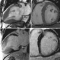
A 32-year-old woman was referred for renal magnetic resonance angiography (MRA) because of suspected renovascular hypertension ( Figure 22-2 ).