Reproductive System


Reproductive System


Female Reproductive System


Anatomy and Physiology
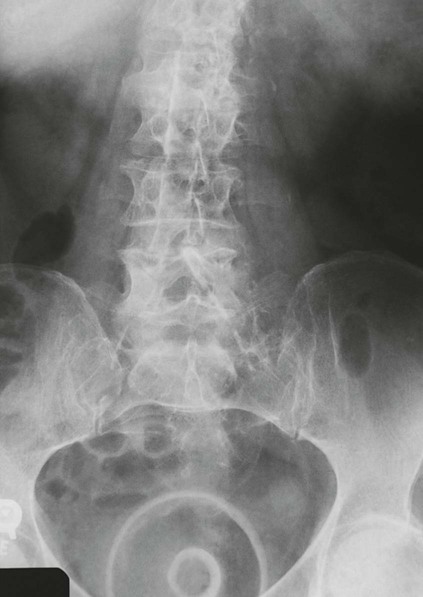

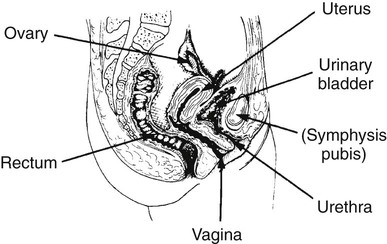
The female reproductive system consists of one pair of ovaries, which are the primary sex organs, and the secondary sex organs, which include one pair of fallopian tubes, the uterus, the vagina, and two breasts (Fig. 10-1). The primary function of the system is to provide a female reproductive cell (the ovum), hormones, and a site for the development of the zygote.


The external genitalia (the vulva) include the mons pubis, the labia majora and minora, the clitoris, the openings of the urethra and vagina, and the perineum. The vagina connects the external genitalia with the uterus and is the mode of exit for menstrual fluids and conception products.


The uterus is a pear-shaped organ, and its primary purpose is to provide an environment for fetal growth and development. Located within the pelvic cavity, it can be divided into the upper portion, termed the fundus; the midportion, termed the body; and the lower portion, termed the cervix. The cervix connects the uterine cavity with the upper vagina. Anatomically, the uterus is flexed so that the cervix and lower portion of the body lie anterior to the rectum. The body of the uterus follows the contour of the posterior urinary bladder wall. The upper portion, or fundus, normally lies superior to the bladder. The walls of the uterus include an inner, endometrial layer; a middle, muscular, myometrial layer; and an outer layer, termed the parietal peritoneum. In actuality, the parietal peritoneum drapes over the fundus and the upper three fourths of the body but does not enclose the lower fourth of the body or the cervix. The actual cavity within the uterus is fairly small and can be well visualized with ultrasonography. It is divided into the internal os, leading to the cervical canal, and the external os, which opens into the vagina. The uterus is held in place within the pelvic cavity via eight ligaments.


Occasionally, lack of proper uterine support is present, and a device known as the pessary (Fig. 10-2) is inserted into the vagina to provide proper support. Current research has demonstrated that the majority of women who use the pessary are very satisfied, as the device relieves the majority of urinary symptoms. Some women report that although the use of a pessary helps improve urinary symptoms, stress incontinence remains an ongoing problem. However, the use of the pessary continues to be the treatment for pelvic organ prolapse and may assist with the quality of life for many of these patients.


The fallopian tubes extend from the upper, outer edges of the uterus and expand distally into the infundibulum located close to, but not attached to, the ovaries. Suspended in place by the broad ligament, they are 8 to 12 cm long and tend to fall behind the uterus. These tubes serve as a passageway for the mature ova and are the normal site of fertilization. In a normal pregnancy, the fertilized ovum continues to travel through the fallopian tube and implants itself into the endometrium of the uterus.


The ovaries are the primary reproductive glands and are responsible for ovulation and for secretion of estrogen and progesterone. Attached to the broad ligament and the posterior uterine wall, each ovary contains numerous graafian follicles enclosing ova. After puberty, several graafian follicles and ova grow and develop each month. Normally only one follicle matures, migrates to the surface of the ovary, and degenerates, thus expelling a mature ovum, which is the hallmark of the ovulation process.


Breasts—like the fallopian tubes, uterus, and vagina—are considered secondary sex organs. Breast parenchyma differs according to age and parity. Women in their 20s and 30s, especially nulliparous women, have dense, fibroglandular parenchyma that may conceal breast masses during a clinical breast examination and imaging. However with advancing age, decline in estrogen and progesterone causes breast tissue to undergo what is known as involutional change. Involution is the conversion of glandular breast tissue into adipose as well as loss of the supportive tissue. As a result of this slow process, the breast changes its architecture, and fibroglandular tissue is replaced by fat. This process is variable, but it typically begins at the back of the breast and progresses forward to the nipple. The term mastalgia applies to conditions that result in pain in one or both breasts. Many patients fear that mastalgia is indicative of breast cancer; however, it is more closely related to architectural changes in breast tissue. Involution aids the imaging professional and the interpreting physician because fatty tissue is radiolucent and enhances the radiographic visibility of many breast masses.


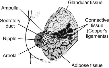
Anatomically, breasts are attached via Cooper ligaments to the pectoral muscles, and this gives the breast its contour and shape. The breast consists of about 12 lobes separated by connective tissue, much like the spokes of a wheel. The lobes are further divided into lobules clustered around small ducts. These small ducts join to form larger ducts, which terminate at the nipple (Fig. 10-3). Breasts function as accessory reproductive glands to secrete milk for the newborn. During pregnancy, changes in estrogen and progesterone levels prepare breasts for lactation. Approximately 3 days after delivery, a lactogenic hormone stimulates the secretion of milk.


Imaging Considerations


Hysterosalpingography
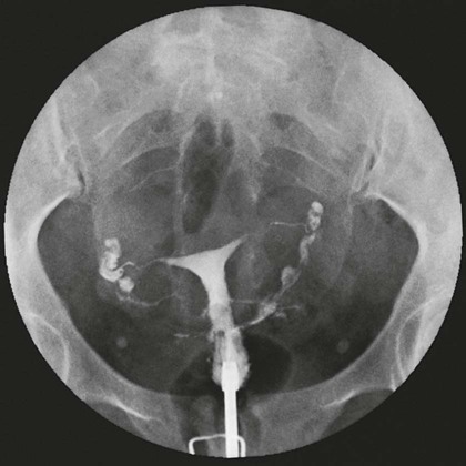

One of the most common radiographic studies of the female reproductive system is the hysterosalpingography (HSG). It is an examination performed for screening of the nongravid (nonpregnant) woman, especially in cases of suspected infertility. A common finding in cases of infertility is nonpatent fallopian tubes. In addition, although HSG does not define the extent of certain conditions such as endometriosis, it is useful in revealing the shape of the uterus and certain characteristics of the fallopian tubes other than their patency. The practice guideline of the American College of Radiology (ACR) for the performance of HSG is to inject approximately 10 to 30 mL of contrast medium into the uterine cavity and the injection should be done slowly to avoid causing spasms and discomfort. Spillage of the contrast medium from the fallopian tubes indicates the patency of the tubes (Fig. 10-4). Typically, HSG is used for diagnostic purposes, but it can also be used therapeutically for restoring tubal patency or to dilate or stretch the fallopian tubes.


An adjunct procedure that has the potential to replace the conventional HSG is known as sonohysterography (SHG). This examination is similar to hysterosalpingography in procedural approach; however, with SHG, normal saline, instead of an iodinated contrast agent, is injected into the uterus. Because saline is devoid of the complications associated with iodinated contrast agents, the procedure may be better tolerated by the patient. In the current application of SHG, saline is used to pry apart the layers of the endometrium to reveal abnormalities within the uterus. The saline fluid is expelled via the fallopian tubes, and this may also indicate their patency. This procedure is viewed via a transvaginal sonographic probe as the physician injects the saline. With real-time images, the dynamics of the reproductive system can be assessed without radiation to the patient.


In a study of a group of 100 patients with infertility, HSG was compared with SHG. These patients’ HSG results were matched to their laparoscopic results. In this small group of women, HSG and SHG were equally effective in the evaluation of tubal disease.


Mammography


The use of mammography as a diagnostic procedure for symptomatic patients is well documented. Mammography provides important information about specific clinical problems such as a breast mass, pain, nipple discharge, and abnormalities of the skin and lymph nodes. With modern mammographic equipment and techniques, radiation exposure is minimal, and evidence does not suggest significant risk to women over 35 years of age. If a risk does exist, it is thought to be so minimal that it has never been observed, but only inferred scientifically.


The use of mammography in asymptomatic patients for screening purposes is based on its ability to detect nonpalpable breast lesions at an early stage when they are too small to be identified by physical examination. Current literature suggests that mammography can detect some cancers 2 years before they are palpable; survival depends on tumor size and lymph node involvement. It is generally agreed that women 50 years of age and older should undergo regular mammographic screening because in this age group, breast tissue is less sensitive to radiation and the incidence of breast cancer increases with age. This also takes advantage of the involutional process, making occult lesions easier to identify on a radiograph. The benefits far outweigh the associated risks from radiation exposure.


Mammography is also a valuable examination tool in the detection and evaluation of breast disease in individuals with augmentation prostheses. Although experience with patients who have undergone augmentation mammoplasty is limited, current research indicates that mammography can demonstrate both palpable and nonpalpable breast lesions. For mammographic screening of these patients, it is important to displace the implant so that the native breast tissue can be imaged and assessed for disease. To demonstrate the underlying breast parenchyma in these individuals, implant-displaced views are required. Technologists are encouraged to use the Eckland maneuver to displace the implant from the native breast tissue to radiograph as much of the native breast tissue as possible. The Eckland maneuver is accomplished by the technologist applying pressure at the area of the nipple and then carefully rolling the native breast tissue away from the implant. The implant-displaced view is performed for both craniocaudal and mediolateral projections of the breast. Once the implant is pushed upward and away from the native breast tissue, the compression paddle is used to continue to hold the implant so that the native breast tissue can be more fully imaged. Because of the variations in patients’ breast tissue and their implants, manual exposure techniques are commonly used.


Needle guidewire localization is a specialized procedure to identify nonpalpable, mammographically detected abnormalities of the breast. It helps direct the surgeon to the lesion in question and allows excision of the suspect tissue for biopsy. Needle guidewire localizations cause minimal morbidity, with complications including hematoma formation, intraoperative wire dislodgement, and wire breakage. The development and refinement of localization techniques have greatly increased the percentage of positive findings on surgical biopsy and allow more accurate diagnosis and treatment of early-stage carcinoma of the breast. Fine-needle and large-core biopsy techniques offer an alternative to surgical biopsy as an initial step in the investigation of breast masses. These procedures are performed on an outpatient basis by the mammographer and the radiologist or by the surgeon in the mammography area using a specially designed stereotactic localization unit. Ductal lavage of the breast may also be performed in cases of suspected intraductal disease to obtain a specimen for laboratory analysis. Use of sonography-guided aspiration and biopsy is also common because sonography provides the ability to visualize the area in question and note whether the area has been completely removed while the needle or biopsy gun is still in situ. This may prevent additional punctures into the patient’s skin during the biopsy procedure.


In patients who have a positive finding on screening mammography, according to the ACR Appropriateness Criteria, diagnostic mammography is rated as highly appropriate as the next step in a patient’s diagnostic workup. The only finding on screening mammography that would not result in the use of diagnostic mammography is a mass that has circumscribed margins, no additional malignant qualities, and has not enlarged compared with the last mammogram. In this case, the use of breast sonography would be highly appropriate.


Sonography


Sonography is the primary modality for examining the gravid or nongravid female reproductive system because of its excellent accuracy and because it presents no radiation hazards to the fetus or the mother. Not only is sonography applicable in pregnancy, but it is also useful in normal gynecologic examinations to visualize the reproductive organs or to monitor the progress of a regimen of fertility medication.


Transabdominal pelvic sonography requires a distended urinary bladder to serve as an “acoustic window” for good visualization of the pelvic organs. In addition, the fluid within the urinary bladder displaces bowel gas away from the area of interest. Sonography of the uterus and ovaries has been greatly enhanced by the use of the transvaginal transducer, which provides more accurate clinical information with magnification of the images of internal pelvic structures. The most common indications for sonography in the nongravid female include evaluation of pelvic, uterine, and ovarian masses because sonography can provide information about mass size, location, internal characteristics, and effect on surrounding organs. Obstetrically, sonography is the method of choice for visualizing the position of the placenta, multiple gestations, or ectopic pregnancies and for determining gestational age. It is used to assist and guide the physician during amniocentesis and is invaluable in assessing fetal abnormalities such as anencephaly, hydrocephaly, congenital heart defects, polycystic kidney disease, urinary tract obstructions, and gastrointestinal (GI) tract obstructions and in determining fetal death.


The use of sonography to assess the pregnant uterus for determining fetal growth, any disturbances, or risk of growth restriction is recommended by the ACR. Sonography is the best imaging modality to assess the fetus, growth, and amniotic fluid and for an overall survey of fetal well-being.


Sonography is an excellent modality for differentiating cystic masses from solid masses within the breast. However, sonography has limitations in the diagnosis of malignant breast disease because of the solid nature of most breast cancers. Recently, researchers and clinicians have made great strides in the use of sonography to evaluate the dense breast for disease. To reduce the radiation dose to young patients, research continues to strive to perfect an accurate and safe screening modality. However, currently, breast sonography is not advocated as a screening modality for breast cancer because of the difficulty in consistently differentiating between a solid benign mass and malignant disease.


Magnetic Resonance Imaging


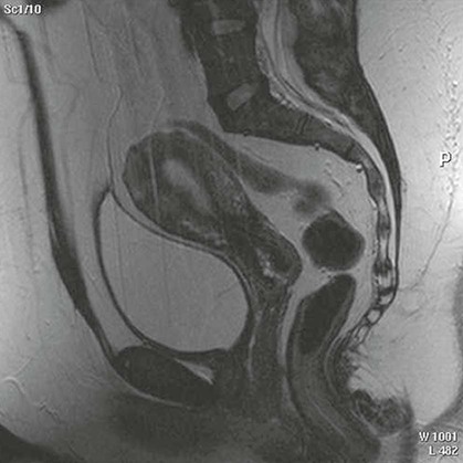
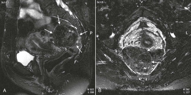
Magnetic resonance imaging (MRI) is now often used in conjunction with sonography in the evaluation of the female pelvis (Figs. 10-5 and 10-6). MRI, like sonography, uses no ionizing radiation and is noninvasive. MRI gives detailed information on pelvic, uterine, and ovarian masses (Fig. 10-7). In cases of ovarian cancer, MRI accurately demonstrates proliferation into other pelvic structures. In addition, multiple leiomyomas can be detected and localized in a short period.


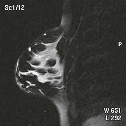
MRI is increasingly being used to assist in differentiating between malignant and benign solid lesions within the breast. Fat suppression imaging is used before and after contrast enhancement. This technique suppresses the normal, fatty tissue of the breast, allowing easier identification of malignant masses through contrast enhancement (Fig. 10-8). MRI is also used to detect faulty or leaking breast implants. Again, fat suppression imaging is used to suppress the normal breast tissue and detect the presence of silicone in surrounding tissue.


A review of published studies on the value of adding MRI as a screening tool for breast cancer found that it was better for screening patients compared with mammography alone or a combination of mammography, sonography, and clinical breast examination. One concern about using MRI as a screening tool for breast cancer was that it raised the return rate for additional imaging; therefore, it has been suggested that MRI be used as an adjunct to mammography. MRI has also proven to be a safe, more effective screening tool in women who have a family history of breast cancer and are positive for the BRCA oncogene, for whom early screening is recommended.


Computed Tomography


Computed tomography (CT) of the pelvis and abdomen is often performed to diagnose diseases of the female and male reproductive systems. It is quite helpful in assessing neoplastic growth and abscess formation resulting from inflammatory processes. It is often used in conjunction with transvaginal sonography to evaluate ovarian lesions, especially cystic teratomas in females, and is used extensively in staging cancers of the female reproductive system. In the male patient, CT is also used in conjunction with sonography to demonstrate anomalies of the seminal vesicles and prostate gland. CT and positron emission tomography (PET) fusion studies (CT-PET) are also of value in the diagnosis and staging of neoplastic disease of the reproductive system, as well as the assessment of disease progression.


Sonography is highly recommended by the ACR for suspected gynecologic adnexal masses. CT of the pelvis, with or without contrast, is also equally indicated for nongravid women in the reproductive age group who have a complex or solid mass that is growing in the short term.


Congenital Anomalies


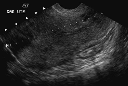
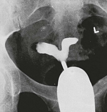
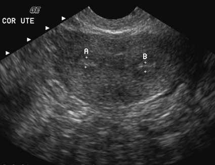
Congenital anomalies of the female reproductive system occur in approximately 1% to 2% of women in the United States. The most common anomaly is the bicornuate uterus, in which paired uterine horns extend into the fallopian tubes (Figs. 10-9 and 10-10). A unicornuate uterus occurs when the uterine cavity is elongated and has a single fallopian tube emerging from it. Often, the kidney on the side of the missing fallopian tube is also absent. Uterus didelphys is a rare congenital anomaly with complete duplication of the uterus, cervix, and vagina. The most serious complication of these anomalies is problems with reproduction, although various surgical corrections can be performed.


In the normal female reproductive system, the fundus of the uterus lies anterior to the cervix and away from the rectum and is said to be anteverted. Occasionally, the normal uterus may lie in an abnormal position. If the uterus is more vertical than normal and points backward toward the bowel, it is said to be retroverted. If the uterus is completely bent back and lies against the rectosigmoid region of the bowel, it is said to be retroflexed. A uterus that is tilted vertically forward is anteflexed, and it lies on top of the urinary bladder. Although neither position is normal, this is generally of little clinical significance.


Mar 6, 2016 | Posted by in GENERAL RADIOLOGY | Comments Off on Reproductive System

Full access? Get Clinical Tree

Get Clinical Tree app for offline access