Resting-state functional MR imaging (rsfMR imaging) measures spontaneous fluctuations in the blood oxygen level–dependent (BOLD) signal and can be used to elucidate the brain’s functional organization. It is used to simultaneously assess multiple distributed resting-state networks. Unlike task-based functional MR imaging, rsfMR imaging does not require task performance. This article presents a brief introduction of rsfMR imaging processing methods followed by a detailed discussion on the use of rsfMR imaging in presurgical planning. Example cases are provided to highlight the strengths and limitations of the technique.
Key points
- •
Resting-state functional MR imaging (rsfMR imaging) is a promising technique for presurgical planning with the objective of decreasing morbidity while maximizing complete resection of pathologic tissue. However, the methodology is still in early stages of development.
- •
Further research is necessary to make these tools more accurate and available in the operating room.
- •
Additional research is needed to explore the differences between rsfMR imaging, task fMR imaging, and electrocortical stimulation mapping, and to better understand the consequences of disrupted resting-state networks outside the motor and language systems.
- •
Related engineering development should incorporate the presurgical MR imaging results into intraoperative neuronavigation systems, including the rsfMR imaging results in conjunction with white matter fiber bundle anatomy derived from diffusion tensor imaging.
Introduction
Background
Functional MR imaging (fMR imaging) detects changes in the blood oxygen level–dependent (BOLD) signal that reflect the neurovascular response to neural activity. Traditionally, fMR imaging has been used to localize function within the brain by presenting a stimulus or imposing a task (eg, presenting a flashing checkerboard pattern or generating verbs from nouns) to elicit neuronal responses. This type of experiment has been very effective at localizing functionality within the brain, as evidenced by the many thousands of publications using task-based fMR imaging.
The human brain consumes a disproportionate amount of energy relative to its weight. The brain constitutes approximately 2% of the body’s weight but consumes 20% of the body’s energy use. Performance of a task only minimally increases energy expenditure. Thus, task-based experiments ignore most of the brain’s activity, which is largely devoted to signaling.
Biswal and colleagues were the first to demonstrate that spontaneous fluctuations in the BOLD signal in the resting state correlated within the somatomotor system. Before this observation, spontaneous fluctuations in the BOLD signal in the resting state were regarded as noise and generally averaged out over many trials or task blocks. More recent studies have shown that these spontaneous fluctuations reflect the brain’s functional organization. Correlated intrinsic activity is currently referred to as functional connectivity MR imaging or resting-state fMR imaging (rsfMR imaging). The development of these methods has opened up many exciting possibilities for future neurocognitive research as well as clinical applications. This article focuses on the application of rsfMR imaging to presurgical planning. Table 1 summarizes key features of both task-based fMR imaging and rsfMR imaging. Snyder and Raichle give a historical review of the resting state.
| Task-Based fMR Imaging | rsfMR Imaging |
|---|---|
| Neuronal activity is studied while performing a well-defined task (eg, finger tapping or object naming). | Neuronal activity is studied in the absence of a task (when the subject in the scanner is in a state of quiet wakefulness). |
| Maps a single functional system at a time | Maps all functional systems simultaneously |
| Task-related changes in the BOLD signal are measured. Regions in the brain associated with a given task are localized. | Spontaneous fluctuations in the BOLD signal are measured. Correlated intrinsic activity defines functional connectivity. |
| Immune to spurious variance in the BOLD signal (eg, P co 2 levels, head motion) | Vulnerable to contamination and requires denoising to remove sources of spurious variance |
Resting-State Networks
Correlated intrinsic activity defines functional connectivity. Functionally connected regions are known as resting-state networks (RSNs); equivalently, intrinsic connectivity networks. The rsfMR imaging scans generally are acquired while the subject is in a state of quiet wakefulness. The importance of RSNs is that their topography closely corresponds to the topography of responses elicited by a wide variety of sensory, motor, and cognitive tasks. Intrinsic activity persists, albeit in somewhat modified form, during sleep or even under sedation. The persistence of the spontaneous fluctuations during states of reduced awareness suggests that intrinsic neuronal activity plays an important role in the maintenance of the brain’s functional integrity. Spontaneous BOLD activity has been detected in all mammalian species investigated thus far, which reinforces the notion that this phenomenon is important from a physiologic and evolutionary point of view. However, the precise physiologic functions of intrinsic activity remain unknown. Examples of important RSNs follow and are summarized in Table 2 .
| 1. Default mode network | Most robust RSN More active at rest than during performance of goal-directed tasks |
| 2. Somatomotor network | Includes primary and higher order motor and sensory areas |
| 3. Auditory network | Includes Heschl gyrus, superior temporal gyrus, and posterior insula |
| 4. Visual network | Includes most of the occipital cortex |
| 5. Language network | Includes Broca, Wernicke, and multiple other language-related areas Extends to prefrontal, temporal, parietal, and subcortical regions |
| 6. Dorsal attention network | Tasks requiring spatial attention Includes intraparietal sulcus and frontal eye field |
| 7. Ventral attention network | Involved in detection of environmentally salient events Includes temporal-parietal junction |
| 8. Frontoparietal control network | Associated with working memory and control of goal-directed behavior Includes lateral prefrontal cortex and inferior parietal lobule |
| 9. Cingulo-opercular network | Associated with performance of tasks requiring executive control Medial superior frontal and anterior prefrontal cortices, anterior insula |
Default mode network
Perhaps the most fundamental RSN is the default mode network (DMN) ( Fig. 1 A), first identified by a meta-analysis of task-based functional neuroimaging experiments performed with positron emission tomography. The defining property of the DMN is that it is more active at rest than during performance of goal-directed tasks. The DMN was first identified using rsfMR imaging by Greicius and colleagues. This finding has since been replicated many times over using a variety of analysis methods. Some investigators have hypothesized that there are two large anticorrelated systems in the brain, one anchored by the DMN and the other composed of systems controlling executive and attentional mechanisms. This dichotomy has been variously referred to as task-positive versus task-negative and intrinsic versus extrinsic. Although the nomenclature associated with the DMN remains controversial, the topography of the DMN is remarkably consistent across diverse analysis strategies.
Sensory and motor resting-state networks
The somatomotor network, first identified by Biswal and colleagues, encompasses primary and higher order motor and sensory areas (see Fig. 1 B). The visual network spans much of the occipital cortex (see Fig. 1 C). The auditory network includes Heschl gyrus, the superior temporal gyrus, and the posterior insula. The language network includes Broca and Wernicke areas but also extends to prefrontal, temporal, parietal, and subcortical regions (see Fig. 1 D).
Attention and cognitive-control resting-state networks
RSNs involved in attentional and cognitive control include the dorsal attention network and the ventral attention network. The dorsal attention network (see Fig. 1 E) includes the intraparietal sulcus and the frontal eye fields and is recruited by tasks requiring control of spatial attention. The ventral attention network (see Fig. 1 F), which includes the temporal-parietal junction and ventral frontal cortex, is involved in the detection of environmentally salient events. The frontoparietal control network (see Fig. 1 G), which includes the lateral prefrontal cortex and the inferior parietal lobule, is associated with working memory and control of goal-directed behavior. Finally, the cingulo-opercular network, also known as the salience network or the core control network, includes the medial superior frontal cortex, anterior insula, and anterior prefrontal cortex. The cingulo-opercular network is thought to enable the performance of tasks requiring executive control.
Application to presurgical planning
Review of the Literature
Multiple studies have demonstrated that maximal resection of a brain tumor while sparing nearby eloquent cortex leads to improved outcomes with reduced morbidity. Similar considerations apply to surgical resections for intractable epilepsy. Historically, neurosurgeons have been concerned with localization of the motor and language system because these parts of the brain instantiate critical functionality (eloquent cortex). However, a broader understanding of brain function suggests that all parts of the brain contribute to important functionality. Thus, improved functional mapping of multiple RSNs beyond motor and language systems could lead to further improvements in patient outcomes.
Several publications have explored the use of rsfMR imaging for use in presurgical planning. An early case report described use of rsfMR imaging to localize the motor cortex in a patient with a brain tumor. Kokkonen and colleagues similarly compared motor task-based fMR imaging data to resting-state data and showed that the motor functional network could be localized based on resting-state data in eight subjects with tumor, as well as 10 healthy control subjects. In surgery for epilepsy, the higher spatial resolution afforded by rsfMR imaging over electroencephalography could provide a distinct advantage in mapping epileptic foci or networks. Seed-based methods were used by Liu and colleagues to successfully locate sensorimotor areas by using rsfMR imaging in subjects with tumors or epileptic foci close to sensorimotor areas. They found agreement between rsfMR imaging and task-based fMR imaging, as well as intraoperative cortical stimulation data. In another study from the same laboratory, Stufflebeam and colleagues were able to localize areas of increased functional connectivity in five of six subjects that overlapped with epileptogenic areas identified by invasive encephalography. Zhang and colleagues used graph methods and a pattern classifier applied to rsfMR imaging data to identify subjects as either having medial temporal lobe epilepsy or as normal controls. Using data from 16 subjects with intractable medial temporal lobe epilepsy and 52 normal controls, they achieved an average classification sensitivity of 77.2% and a specificity of 83.86%. Bettus and colleagues reported that increases in basal functional connectivity were a specific marker of the location of the epileptogenic zone in 22 subjects with mesial temporal lobe epilepsy. Weaver and colleagues studied four subjects with nonlesion, focal epilepsy along with 16 control subjects to determine whether the seizure focus could be found using the functional patterns near the epileptogenic zone. By averaging voxel homogeneity across regions of interest and comparing that with other regions, they were able to accurately identify the epileptic focus. Tie and colleagues used spatial independent component analysis (sICA) on a training group of 14 healthy subjects to identify the language network based on rsfMR imaging. The result of that analysis was then used to identify the language network in a second group of 18 healthy subjects at the individual level. They further proposed an automated system for localizing the language network in individual patients using sICA. A more detailed presentation of the authors’ experiences using rsfMR imaging for presurgical mapping with both seed-based and multilayer perceptron (MLP) approaches follows.
Overview of Processing Methods
rsfMR imaging methodology currently is dominated by two complementary strategies: spatial sICA and seed-based correlation mapping. Both strategies depend on spontaneous neural activity being correlated (phase coherent) within widely distributed regions of the brain. Both strategies yield highly reproducible results at the group level. sICA decomposes rsfMR imaging data into a sum of components, each component corresponding to a spatial topography and a time course. In contrast, seed-based correlation mapping is computed by voxel-wise evaluation of the Pearson correlation between the time courses in a targeted region of interest and all other voxels in the brain.
The principal advantage of sICA is that it provides a direct means of separating artifact from BOLD signals of neural origin, although this separation typically requires observer expertise. The results obtained using sICA may vary substantially depending on processing parameters (eg, number of requested components). Thus, sICA can be difficult to use in the investigation of targeted RSNs, especially in single subjects. In contrast, targeting of selected RSNs is built into seed-based correlation mapping. However, the principal difficulty in using seed-based correlation mapping is exclusion of nonneural artifact, which is typically accomplished using regression techniques.
sICA and seed-based correlation mapping both represent strategies for assigning RSN identities to brain voxels. Because sICA makes no a priori assumptions regarding the topography of the obtained components, this method exemplifies unsupervised classification. In contrast, seed-based correlation mapping depends on previous knowledge, and so exemplifies supervised classification. Hacker and colleagues discuss the distinction between supervised and unsupervised methodologies.
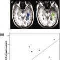
Hacker and colleagues recently described a technique for mapping the topography of known RSNs in individuals using MLP. Perceptrons are machine learning algorithms that can be trained to associate arbitrary input patterns with discrete output labels. An MLP was trained to associate seed-based correlation maps with particular RSNs. Running the trained MLP on correlation maps corresponding to all voxels in the brain generates voxel-wise RSN membership estimates. Thus, RSN mapping using a trained MLP exemplifies supervised classification. An example of the RSN produced by the MLP algorithm in three subjects is presented in Fig. 2 .
Related posts:
 Clinical Applications of Functional MRI
 Foreword
 Blood Oxygen Level Dependent Functional Magnetic Resonance Imaging for Presurgical Planning
Technical Considerations for Functional Magnetic Resonance Imaging Analysis
Blood Oxygen Level Dependent Functional Magnetic Resonance Imaging for Presurgical Planning
Technical Considerations for Functional Magnetic Resonance Imaging Analysis
 Pretherapeutic Functional Magnetic Resonance Imaging in Children
 Applications of Blood-Oxygen-Level-Dependent Functional Magnetic Resonance Imaging and Diffusion Tensor Imaging in Epilepsy
Stay updated, free articles. Join our Telegram channel

Full access? Get Clinical Tree


