Location: R. of the limbs constitutes 15 % of all tumors. Type (a): Head, neck, genitourinary tract (botryoid variety), retroperitoneum. Type (b): Limbs (forearm, hand, foot). Type (c): Limbs (prevails in the thigh).
Clinical: Type a): Rapidly, invasive, infiltrative growth. Symptoms and signs depend on anatomic location. Types (b–c): Deep, painless unless it compresses a nerve, rapidly enlarging mass in a striated muscle.
Imaging: On x-ray erosion of flat bone in 20 % of cases. In types (b–c) is rare, but it occurs mainly in the hand and foot. Periosteal reaction is minimal. Nevertheless, tumor is frequently tightly bound to bone. On arteriography common aspects to all well-vascularized tumors: neovascularity and tumoral blush. On CT large, lobulated mass with central hypodensities, septation, rim-like enhancement surrounding areas of necrosis after contrast administration. On MRI type (a): homogeneous, slight hyperintensity on T1, bright on T2 with some dark internal strands, no enhancement of large parts of the mass on contrast T1, no necrosis. Type (b): Inhomogeneous, intermediate intensity with multiple, central hypointensity areas on T1, heterogeneous, with white central zones on T2 (necrotic areas), strong peripheral enhancement on contrast T1. Type (c): Mixed signal intensities on T1, more inhomogeneous on T2 with central fluid collection near to low-signal-intensity areas, ringlike, peripheral enhancement around these.
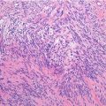
Histopathology: Firm or soft mass, with myxoid and cystic component, a mottled white-gray to pink-tan, with frequent hemorrhage and necrotic areas. Botryoid variety has a polypoid and myxoid appearance compared to a bunch of grapes. Immunostains for desmin and myoglobin are essential for diagnosis. Type (a): Usually, undifferentiated, small, round cells with round-oval, hyperchromic nucleus. Nuclear membrane is evident. Chromatin granules are distinct, and nucleoli are clearly evident. Thin, perinuclear cytoplasmic ring. Mitotic figures and pleomorphism are frequent. Cells are distributed without any order with scarce collagen stroma among them. At other times, in more differentiated zones, the volume of the cells increases, and the nucleus becomes larger, vesicular, and eccentric, with a large nucleolus. Cytoplasm is eosinophilic, granular, and more abundant, with filaments wrapped around the nucleus. Transverse striation may be recognized. Racket or tadpole or ribbon shapes, angulated like broken straws of hay, are observed. Globose cells may have a vacuolated cytoplasm, and this may be reduced to filaments that from the nucleus irradiate to the periphery: spider-web cells. Type (b): Undifferentiated, small, round-oval cells, collected in solid islands and forming alveolar aspect. Rough bands of dense collagen, convoluted and ramified, containing dilated capillaries, surround these cellular zones. Peripheral cells adhere in a single layer to the fibrous septa. Cells in the center are more loosely arranged or freely floating. Rhabdomyoblasts are much rarer. Type (c): Association of loosely arranged, haphazardly oriented, small and large, round or pleomorphic cells. Larger and more irregular in outline racket-shaped and tadpole-shaped rhabdomyoblasts with stringy, granular, and vacuolated cytoplasm. Transverse striation is extremely rare. Much less collagen stroma between cells is observed. Immunohistochemistry is positive for vimentin, desmin, pan-muscle actin, MyoD1, and myogenin (diffuse positivity in type (b) but negative for smooth cell actin. There are two main karyotypic aberrations in alveolar rhabdomyosarcoma: t(2;13) that fuses the PAX3 gene on 2q35 with the FOXOA1 gene on 13q14 and is present in 60–70 % of cases and t(1;13) that fuses PAX7 on 1p36 with FOXOA1 on 13q14 and is present in 10–20 % of cases. The latter translocation is associated with a better prognosis, the first with a worse prognosis.
Related posts:


Stay updated, free articles. Join our Telegram channel

Full access? Get Clinical Tree


