Rheumatoid Necrobiotic Nodules
Helen T. Winer-Muram, MD
Key Facts
Terminology
Subacute or chronic inflammatory polyarthropathy of unknown cause
Imaging Findings
Rheumatoid nodules (seen in < 5%)
Solitary or multiple, 0.5-7 cm, few in number
Peripheral (subpleural)
Cavitation (50%)
May contain necrotic lung ball or rarely mycetoma
May result in pneumothorax (may be refractory to therapy)
Wax & wane
Caplan syndrome: Rare
Hypersensitivity reaction to dust
Associated with coal miner’s pneumoconiosis
Large rounded nodules (0.5-5 cm), may cavitate
Rheumatoid arthritis nodules may have uptake on PET
Top Differential Diagnoses
Tuberculosis
Histoplasmosis
Metastases
Amyloid Nodules
Pathology
Nodules identical to subcutaneous nodules
Clinical Issues
Rheumatoid nodules, more common in males, especially smokers
Diagnostic Checklist
Need to rule out infectious etiology for nodules prior to initiation of immunosuppression
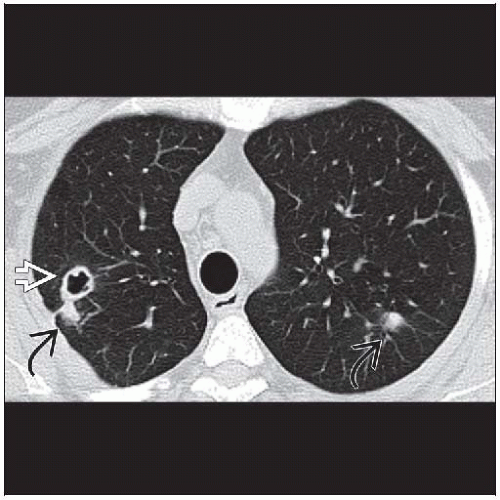 Axial NECT shows multiple solid  and cavitary and cavitary  pulmonary nodules from rheumatoid arthritis. pulmonary nodules from rheumatoid arthritis. |
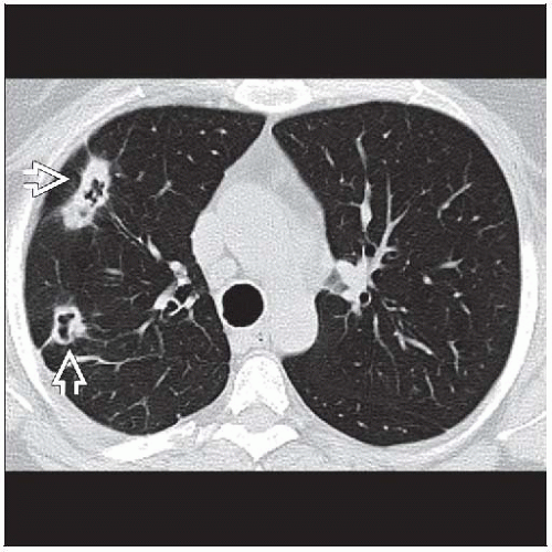 Axial NECT in the same patient shows large peripheral cavitary nodules with variable wall thickness  from rheumatoid necrobiotic nodules. from rheumatoid necrobiotic nodules. |
TERMINOLOGY
Abbreviations and Synonyms
Rheumatoid arthritis (RA)
Caplan syndrome, rheumatoid pneumoconiosis
Definitions
Subacute or chronic inflammatory polyarthropathy of unknown cause
IMAGING FINDINGS
General Features
Best diagnostic clue: Chronic pleural effusion and lung nodules in patient with rheumatoid arthritis
Patient position/location: Peripheral subpleural
Size: Variable, usually < 1 cm diameter
Morphology: Solid or cavitary nodules
CT Findings
Rheumatoid nodules (seen in < 5%)
Morphology
Solitary or multiple, 0.5-7 cm, few in number
Usually well circumscribed
Resemble neoplasm, discrete, rounded, or lobulated
May rarely calcify
Identical to subcutaneous nodules (usually seen around elbows)
Cavitation (50%)
Thick or thin smooth wall
May contain necrotic lung ball or rarely mycetoma
May result in pneumothorax (may be refractory to therapy)
Distribution
Peripheral (subpleural)
Evolution
Wax & wane
Caplan syndrome: Rare
Hypersensitivity reaction to dust
Associated with coal miner’s pneumoconiosis
Redefined to include silica, asbestos, dolomite, carbon
Large rounded nodules (0.5-5 cm), may cavitate
Indistinguishable from silicate pneumoconiotic nodules or tuberculous nodules
Nodules have peripheral distribution
Serologic but not clinical rheumatoid arthritis
Other associated pleuropulmonary abnormalities
Pleural disease
Most common abnormality in RA
Pleural effusions or thickening from fibrothorax
Effusions small to large
Effusions usually unilateral, transient, persistent, or relapsing
May be associated with rheumatoid nodules and subcutaneous nodules
Pleural abnormalities and pulmonary nodules, if present, help distinguish RA-related interstitial lung disease
Interstitial pneumonitis & fibrosis
Most common pulmonary abnormality (30-40%)
Nonspecific interstitial pneumonia or usual interstitial pneumonia
Cryptogenic organizing pneumonia pattern
Ground-glass opacities, nodules, bronchovascular distribution
Airways disease
Bronchiectasis, follicular bronchiolitis, constrictive bronchiolitis
Radiographic Findings
Nodules may appear in crops, persist or resolve spontaneously, wax & wane
Serial radiography for surveillance
Development of new nodules
Stability or resolution of nodules
Progression: Increasing size and number of nodules, development of cavitation
Development of pleuropulmonary complications
Imaging Recommendations
Best imaging tool: HRCT useful to characterize pattern and extent of diseaseRelated posts:

Stay updated, free articles. Join our Telegram channel

Full access? Get Clinical Tree


