Anatomy, embryology, pathophysiology
- ◼
There are three major anatomic regions that may contribute to right upper quadrant pain. Organ systems with differential diagnoses include:
- ◼
Gastrointestinal system: liver, biliary, pancreas, and duodenum.
- ◼
Liver.
- ◼
Hepatic steatosis.
- ◼
Infectious hepatitis.
- ◼
Alcoholic hepatitis.
- ◼
Autoimmune hepatitis.
- ◼
Toxin-related hepatitis.
- ◼
Hepatic abscess.
- ◼
Portal vein thrombosis.
- ◼
Budd-Chiari syndrome.
- ◼
Perihepatic inflammation (Fitz-Hugh-Curtis syndrome).
- ◼
- ◼
Biliary
- ◼
Cholelithiasis.
- ◼
Acute or chronic cholecystitis.
- ◼
Gallbladder sludge.
- ◼
Choledocholithiasis.
- ◼
Cholangitis.
- ◼
- ◼
Pancreas.
- ◼
Acute or chronic pancreatitis.
- ◼
- ◼
Duodenum.
- ◼
Ulcers.
- ◼
Obstruction.
- ◼
Perforation.
- ◼
- ◼
Right lung base.
- ◼
Pneumonia.
- ◼
Pleural effusion.
- ◼
Pulmonary embolism.
- ◼
- ◼
Musculoskeletal system: right lower ribs, right anterolateral abdominal wall muscles.
- ◼
Muscle strain/costochondritis.
- ◼
Intramuscular hematoma.
- ◼
Herpes zoster.
- ◼
Rib fracture.
- ◼
- ◼
Techniques
Radiography of the abdomen is of limited value for evaluating right upper quadrant pain, although it may identify gallstones.
- ◼
According to the American College of Radiology Appropriateness Criteria, ultrasound remains the initial test of choice for imaging patients with right upper quadrant pain because of its greater availability, shorter study time for identification or exclusion of diagnoses, and lack of ionizing radiation.
- ◼
If ultrasound results are negative, computed tomography (CT) abdomen and pelvis with contrast is the preferred next imaging modality of choice.
- ◼
Magnetic resonance imaging (MRI) abdomen with and without contrast is another alternative for right upper quadrant pain. Although factors, such as longer acquisition times limit its use in the emergency setting, lack of ionizing radiation makes it a preferred choice in young patients over CT.
- ◼
For pregnant patients, ultrasound is the initial imaging test of choice for evaluating right upper quadrant pain. MRI is the preferred test to follow an inconclusive ultrasound.
Protocols
Ultrasound
- ◼
Ultrasonography is a user-dependent modality that allows the operator more freedom when creating representative images.
- ◼
Body habitus and dense tissues can create imaging obstacles for sonographers. The more experienced sonographer can optimize the images by adjusting the ultrasound machine settings and changing transducers to optimize an image.
- ◼
Harmonic imaging technique fused in the gray-scale ultrasound image will improve image conspicuity by decreasing grating lobes, side lobes, and reverberation artifacts.
Computed tomography
- ◼
Several studies have looked at the use of oral contrast media in patients who present to an emergency department with acute abdomen or blunt trauma and found that using oral contrast media did not substantially improve diagnostic performance.
- ◼
Because there are many disadvantages to the use of oral contrast media in these patients, including time delay, inability of patients to ingest large volumes of fluid, and potential need for emergency surgery, many institutions do not use oral contrast agents unless there is suspicion of postoperative bowel leak.
- ◼
Low osmolar contrast media is used because of a lower incidence of adverse reactions.
- ◼
Contrast-induced nephropathy can be prevented by judicious hydration of at-risk patients with low estimated glomerular filtration rate.
- ◼
Patients at high risk of allergic reactions should be premedicated.
Magnetic resonance imaging
- ◼
Patients must be screened for risk factors or contraindications to MRI via comprehensive clinical evaluation and history before imaging.
- ◼
Proper breath-hold instructions must be clearly stated to obtain the highest quality images.
Specific disease processes
Hepatic causes of acute right upper quadrant pain
Acute hepatitis
The imaging findings of the different causes of acute hepatitis are nonspecific and overlap. Hepatomegaly and periportal edema are common findings.
Ultrasound
- ◼
Hypoechoic parenchyma with increased prominence of the portal triad (starry-sky appearance) ( Fig. 10.1 ).
- ◼
Heterogeneous echotexture, gallbladder wall thickening, and accentuation of the portal triads.
Computed tomography
- ◼
Findings are nonspecific but include hepatomegaly, gallbladder wall thickening, and periportal edema, which manifests as low attenuation regions along the portal triads ( Fig. 10.2 ).
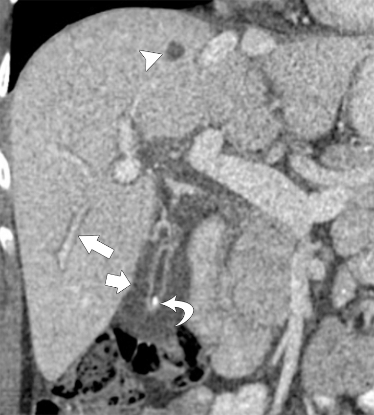
Fig. 10.2
Coronal contrast-enhanced computed tomography in a 36-year-old with acute viral hepatitis, mild hepatomegaly, portal tract edema ( arrow ), and diffuse gallbladder wall thickening ( small arrow ). An incidental simple hepatic cyst ( arrowhead ) and gallstone can be seen ( curved arrow ).
(From Boland GW. Gastrointestinal Imaging: the Requisites , ed 4. Philadelphia: Saunders; 2014.)
- ◼
After the intravenous injection of contrast material, the liver parenchyma may enhance heterogeneously.
Hepatic abscess
Hepatic infections leading to abscess formation include pyogenic infection, amebiasis, and fungal and parasitic diseases.
Ultrasound
- ◼
Complex cystic lesion with thick irregular wall, septations, and internal echoes.
- ◼
Markedly increased echogenicity with associated reverberation artifact suggests the presence of gas in the lesion.
Computed tomography
- ◼
Single or multiloculated, hypoattenuating lesions with thick enhancing rim (double target sign) ( Fig. 10.3 ).

Fig. 10.3
Computed tomography (CT) findings of pyogenic abscess: transverse contrast-enhanced CT image shows a round, well defined, hypoattenuating lesion in the right hepatic lobe with thick peripherally enhancing capsule.
Portal vein thrombosis
Ultrasound
- ◼
Dilated portal vein with absence of flow.
- ◼
Acute clot is hypoechoic, whereas chronic clot is hyperechoic.
Computed tomography/magnetic resonance
- ◼
Acute thrombosis:
- ◼
Expansion and peripheral enhancement of the vein ( Fig. 10.4 ).

Fig. 10.4
Acute portal vein thrombosis. Axial (A) and coronal (B) contrast-enhanced computed tomography images demonstrating hypoattenuation, expansion, and peripheral enhancement of the portal vein ( arrows in A and B).
- ◼
Associated perfusional abnormality in the liver parenchyma with foci of increased enhancement on the arterial phase and decreased enhancement in the portal venous phase.
- ◼
- ◼
Subacute and chronic thrombosis:
- ◼
Manifests as cavernous transformation of the portal vein, which refers to multiple tortuous venous collaterals at the porta hepatis that replace the portal vein ( Fig. 10.5 ).
- ◼