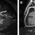
Vascular rings and pulmonary artery slings are rare congenital anomalies that often present with symptoms of tracheal and esophageal compression. These can involve the aortic arch branches and pulmonary arteries, respectively. This review illustrates the current role of MR imaging, highlights its advantages, and provides insight into the diagnosis of these anomalies by describing the embryology and characteristic imaging features of these lesions. Vascular rings and pulmonary artery slings present with symptoms of tracheal and esophageal compression during infancy and childhood. Double aortic arch and right aortic arch with aberrant left subclavian artery and left patent ductus arteriosus (PDA) are the 2 most common types of vascular ring. Atretic segments cannot be directly visualized on MR imaging, but their presence can be inferred from other indirect imaging signs. MR imaging/magnetic resonance angiography (MRA) can diagnose these vascular anomalies and evaluate the airway and esophagus without the use of ionizing radiation. Vascular rings and pulmonary artery slings are rare congenital anomalies of the aortic arch branches or pulmonary arteries that often present with symptoms of tracheal or esophageal compression during childhood. Diagnosis of these conditions can be made using various imaging modalities, including radiography, esophagography, echocardiography, CT, and MR imaging. The purpose of this review is to highlight the advantages of MR imaging for the diagnosis of vascular rings and pulmonary artery slings and to provide an insight into the diagnosis of specific vascular ring types using case examples. MR imaging possesses unique advantages over other imaging modalities in the diagnosis of vascular rings and pulmonary artery slings. Chest radiography can detect a right aortic arch or tracheal compression, and barium esophagography may demonstrate esophageal compression, but these modalities do not directly image vascular structures and cannot provide conclusive anatomic diagnosis, which can be essential for surgical planning. Echocardiography can diagnose aortic arch sidedness and branching pattern, double aortic arches, and pulmonary artery anatomy. Tracheal and esophageal compression cannot be evaluated, however, and due to suboptimal acoustic windows may limit diagnostic accuracy for vascular rings. Angiography can image patent vascular structures but cannot demonstrate tracheal and esophageal compression, requires invasive vascular access with a potential for complications, and requires ionizing radiation and nephrotoxic contrast agent. Due to the ability to delineate vascular anatomy with multiplanar and 3-D reconstruction, and simultaneously demonstrate tracheal and esophageal compression, CT and MR imaging have become the preferred imaging modalities for the diagnosis of vascular rings and pulmonary artery slings. MRA can demonstrate vascular anatomy similar to CT without using ionizing radiation, which is particularly desirable in this young population. MR imaging protocol for vascular ring and pulmonary artery sling evaluation typically consists of gadolinium-enhanced angiography and ECG-gated black blood and bright blood sequences. Black blood imaging in axial and oblique coronal (oriented along the trachea) planes is useful for assessment of vascular anatomy, tracheobronchial tree, and esophagus. Unless contraindicated, 3-D gadolinium-enhanced MRA provides comprehensive assessment of vascular anatomy. ECG and respiratory navigator-gated 3-D steady-state free precession (SSFP) imaging can simultaneously assess vascular, airway, and esophageal anatomy in a 3-D data set without the need for gadolinium. A vascular ring is defined as an abnormality of the aortic arch, its branches, or remnants that results in encircling of the trachea and esophagus with variable degrees of compression. Vascular rings represent 1% to 3% of congenital cardiac anomalies and may remain asymptomatic or present at various times throughout childhood with stridor, dyspnea, chronic cough, wheezing, recurrent respiratory infection, or dysphagia, which may be relieved by surgery. Normal aortic arch development involves variable regression of 6 paired aortic arches from the aortic sac to the paired dorsal aortae. The first, second, and third arches form parts of the maxillary, stapedial, and common carotid arteries, respectively. Remnants of the fourth arch form the distal left aortic arch and proximal right subclavian artery. The fifth arches usually regress, and the sixth arches form parts of the pulmonary arteries and distal ductus arteriosus. The spectrum of aortic arch anomalies and vascular rings can be easily understood using the Edwards hypothetical double arch system ( Fig. 1 ). Normal left arch anatomy occurs after involution of the distal right fourth arch and regression of the right ductus ( Fig. 2 ). Abnormal patterns of regression of these paired aortic arches can result in a vascular ring. Aortic arch sidedness is defined by the location of the aortic arch in relation to the trachea (left, right, or double) as it passes over the mainstem bronchus ( Fig. 3 ). On echocardiography and angiography, this determination is indirect and may be inaccurate. MR imaging directly visualizes both the aorta and tracheobronchial tree, thus allowing conclusive diagnosis. The spectrum of vascular rings ( Table 1 ), with their distinctive MR imaging appearance, is described later.
Key points
Introduction
Advantages of MR imaging
MR imaging protocol
Vascular rings
Normal Aortic Arch Development


Vascular Ring Type
Total Vascular Rings (%)
Association with CHD or Syndromes
Typical Age at Presentation
DAA
50
Usually isolated anomaly
Birth–childhood
Right dominant
70–75 of DAA
Left dominant
20 of DAA
Codominant
5 of DAA
Right aortic arch with aberrant left subclavian
30
Usually isolated anomaly
Infancy–adulthood
May remain asymptomatic due to loose ring
Right aortic arch with mirror image branching and retroesophageal ductus arteriosus
5
Commonly associated with other CHDs, such as tetralogy of Fallot, truncus arteriosus, ventricular septal defect
Infancy–childhood
Circumflex aortic arch
Rare
50%–60% Associated with CHDs, such as ventricular septal defect, double-outlet right ventricle, and coarctation
Birth–childhood Related posts:
![]()
Stay updated, free articles. Join our Telegram channel

Full access? Get Clinical Tree