41 Saphenous Nerve Block
The saphenous nerve is a sensory branch of the femoral nerve that travels with the superficial femoral artery in the thigh along with the infrapatellar nerve and a nerve to the vastus medialis.1 Under the sartorius muscle, this complex forms the subsartorial plexus, which also can include contributions from the posterior division of the obturator nerve.2 The femoral artery and these nerves separate at the entrance to the adductor canal.3,4
The saphenous nerve emerges into the subcutaneous tissue between the sartorius and gracilis tendons within the pes anserinus to join the undersurface of the saphenous vein near the knee crease. The saphenous nerve and vein travel together in the leg to lie anterior to the medial malleolus. Saphenous nerve block is important for surgical anesthesia of the foot and ankle because it innervates the medial leg, malleolus, and foot. There are many approaches to saphenous nerve block along the course of the nerve.5
Suggested Technique
Subsartorial Plexus Block (Mid-Thigh)
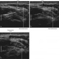
Saphenous nerve block can be performed in the mid-thigh with the patient in the supine position. An in-plane approach from the anterior thigh can be used to direct the needle tip through the sartorius muscle. The nerves of the subsartorial plexus can be imaged as a “bright triangle” anterior to the superficial femoral artery. The needle tip crosses the fascial plane on the anterior side of the nerves to avoid the neurovascular bundle. This subsartorial saphenous block is the deepest saphenous nerve block and therefore requires a steep angle of approach. However, the subsartorial plexus can often be directly imaged with ultrasound. The best access to the fascial layer of the nerves of the subsartorial plexus is usually attained by passing the block needle through the sartorius muscle rather than the vastus medialis. This trans-sartorial approach is similar to previous descriptions of saphenous nerve block that involved nerve stimulation.6
Vastoadductor Membrane Block (Distal Thigh)
Saphenous nerve block also can be performed in the distal thigh with the patient in the supine position.7 An in-plane approach can be used to direct the needle tip through the vastus medialis just adjacent to the sartorius muscle. The muscles are viewed in short axis by placing the transducer on the distal aspect of the medial thigh with the needle introduced anteriorly. The local anesthetic injection should track within the fascial plane deep to the sartorius. Direct imaging of the saphenous nerve and the accompanying branch of the femoral artery is sometimes possible within this fascial plane.
Subcutaneous Infiltration Block (Proximal Leg)
Saphenous nerve block can also be performed in the leg at the level of the tibial tuberosity.8 For this procedure, local anesthetic is infiltrated within the subcutaneous tissue near the saphenous vein using either an in-plane or out-of-plane approach. At this level the saphenous nerve and saphenous vein both lie superficial to the fascia lata. If the saphenous vein is difficult to visualize, a proximal tourniquet can be applied.
Key Points
| Saphenous Nerve Block | The Essentials |
|---|---|
| Anatomy | The SaN lies under the Sa anterior to the SFA. |