Fig. 30.1
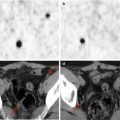
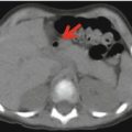
Maximum intensity projection 18F-FDG–PET image (a) shows multiple areas of increased radiopharmaceutical uptake in the thoracic region (arrows). Axial CT (b), PET (c), and PET/CT (d) images show the presence of increased radiopharmaceutical uptake corresponding to several lymph nodes located bilaterally in the mediastinum and in the pulmonary hilar region
Teaching Point
Sarcoidosis typically causes increased 18F-FDG uptake. Thus, in differentiating benign from malignant abnormalities, positive 18F-FDG–PET findings should be interpreted with caution. 18F-FDG–PET/CT is a very useful molecular imaging method in assessing disease activity and in identifying the occult sites of disease in patients with sarcoidosis, including pediatric patients.
Case 2
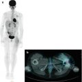
One year before presenting to our clinic, a 17-year-old boy without a remarkable disease history had an EBV infection, with the appearance of lymph nodes in the left lateral cervical region and, on his right side, in the trochlear area. Concurrently, he reported occasional skeletal pain and swelling in the right knee and both feet, progressive rhinitis with anosmia, and polydipsia–polyuria. Due to the persistence of a fever of unknown origin, the patient underwent a 18F-FDG–PET/CT which showed multiple areas of increased tracer uptake in the body (Figs. 30.2, 30.3, 30.4, and 30.5). Histology on some of the 18F-FDG-avid lesions demonstrated granulomatous disease compatible with sarcoidosis. The patient underwent immunosuppressive therapy. A repeated 18F-FDG–PET/CT demonstrated an excellent response to the treatment (Fig. 30.6).
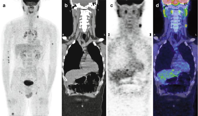
Fig. 30.2


Maximum intensity projection PET image (a) showing 18F-FDG uptake in the lymph nodes of the left laterocervical region, arms, mediastinum, pulmonary hilar and inguinal regions, and in the right leg. Coronal CT with mediastinal window (b), PET (c), and PET/CT fusion (d) images show a 18F-FDG-avid left laterocervical lymph node
Related posts:


Stay updated, free articles. Join our Telegram channel
Full access? Get Clinical Tree
