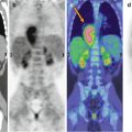
Fig. 22.1
Transaxial FDG–PET scan slices show a hypermetabolic focus in the right temporal mesial cortex (black arrow) and hypometabolism in the ipsilateral lateral and posterior temporal cortex (intra-hemispheric diaschisis, red arrows)
The EEG recorded during the 30 min of FDG uptake during the PET scan showed repeated episodes of the above-described abnormalities (Fig. 22.1). Accordingly, the brain FDG–PET scan was considered as “ictal.” The PET-derived information shown in Fig. 22.1 supported the EEG evidence and helped to guide surgery.
Teaching Point
In patients who are candidates for the surgical treatment of epilepsy and whose MRI study is inconclusive, the brain 18F-FDG–PET may provide relevant information about the site and side of the epileptic focus. Simultaneous EEG recording during the FDG distribution time is mandatory. The epileptic focus will appear as a hypermetabolic site in the unusual case of ictal PET (as in the case described above) and as a hypometabolic area during an interictal (i.e., in the absence of a sustained epileptic discharge as seen on EEG) acquisition.
References
Related posts:


Stay updated, free articles. Join our Telegram channel

Full access? Get Clinical Tree


