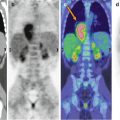
Fig. 29.1
Maximum intensity projection 18F-FDG–PET image (a) shows multiple areas of increased radiopharmaceutical uptake in the thoracic region and in the upper abdomen (arrows). The axial CT (b), PET (c), and PET/CT (d) images show increased radiopharmaceutical uptake corresponding to pulmonary (b–d, first row), lymph node (b–d, second row), and liver (b–d, third row) lesions (arrows). These findings were suspicious for malignancy but histology demonstrated the presence of granulomatous disease and laboratory data suggested a TBC infection
Teaching Point
Mycobacteriosis (including TBC) frequently causes an increased 18F-FDG uptake in affected organs. Thus, in geographic regions with a high prevalence of granulomatous diseases, positive 18F-FDG–PET results should be interpreted with caution in differentiating benign from malignant abnormalities.
Related posts:


Stay updated, free articles. Join our Telegram channel

Full access? Get Clinical Tree


