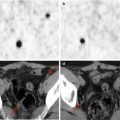
Fig. 31.1
A 10-year-old girl with neurofibromatosis type 1. The plexiform neurofibroma involved the right cervical and axillary region. (a) Axial PET/CT study and (b) axial PET/CT control after 2 years (March 2010) show a mild nonhomogeneous 18F-FDG uptake (SUVmax 1.7) with a focal much intense radiotracer accumulation (SUVmax 3.9)
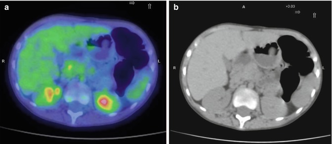
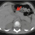
Fig. 31.2
A 9-year-old girl with neurofibromatosis type I and multiple neurofibromas extending from the mediastinum to the cardias. In abdomen the neurofibromas enclose the celiac trunk reaching the porta hepatis. (a) Axial PET/CT and (b) CT images show a mass surrounding the celiac trunk (SUVmax 1.8)
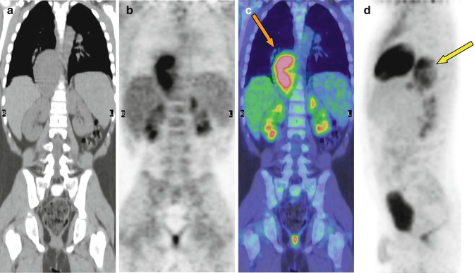
Fig. 31.3
A 7-year-old boy with neurofibromatosis type I. (a) Coronal CT, (b) PET, (c) PET/CT fusion, and (d) sagittal maximum intensity projection PET images show increased FDG uptake corresponding to a paravertebral mass in the left posterior mediastinum (orange arrow in c and yellow arrow in d) between seventh and tenth dorsal vertebrae (SUVmax 9.5). Histology demonstrated the presence of a malignant nerve sheath tumor
Related posts:


Stay updated, free articles. Join our Telegram channel

Full access? Get Clinical Tree


