Sarcoidosis


Sarcoidosis


W. Richard Webb


Sarcoidosis is a systemic disorder of unknown cause, characterized by the presence of noncaseating granulomas. It may involve almost any organ, but most morbidity and mortality result from pulmonary disease. Pulmonary manifestations are present in 90% of patients. Approximately 25% of patients have respiratory symptoms at diagnosis, usually dyspnea; also common are weight loss, fatigue, and sometimes fever or night sweats. Erythema nodosum is common.

Pulmonary lesions may resolve spontaneously or progress to fibrosis; 20% to 25% of patients have permanent functional impairment. Hilar and mediastinal lymph node enlargement is a common finding.


STAGING OF SARCOIDOSIS BASED ON PLAIN RADIOGRAPHS

Sarcoidosis has been described as occurring in stages based on plain radiographic findings:

Stage 0: No visible abnormalities (10% of cases)

Stage 1: Hilar or mediastinal lymph node enlargement not associated with visible lung disease (50% of cases)

Stage 2: Hilar or mediastinal lymph node enlargement associated with visible lung disease (30% of cases)

Stage 3: Diffuse lung disease without lymph node enlargement (10% of cases)

Stage 4: This designation is sometimes used to refer to endstage fibrosis.

The utility of this staging system is limited, although there is some correlation between the stage and the course of disease. Radiographic abnormalities resolve in 65% of stage 1 patients, 50% of stage 2 patients, and 20% of stage 3 patients. However, patients in one stage need not progress to the next. This staging system is not used with CT, which is much more sensitive than radiographs in detecting both lymph node enlargement and lung disease.


LYMPH NODE ABNORMALITIES

Mediastinal lymph node enlargement is very common with sarcoidosis, occurring in 60% to 90% of patients at some stage in their disease. Less than half of patients with lymph node enlargement also show findings of lung disease on plain radiographs. A greater percentage of patients with lymph node enlargement show evidence of lung disease on CT.

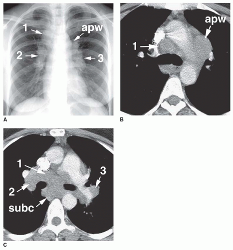
Typically, node enlargement involves the hilar and mediastinal nodes, and lymph node masses usually appear bilateral and symmetrical on chest radiographs. The combination of (1) right paratracheal, (2) right hilar, and (3) left hilar node enlargement, termed the 1-2-3 pattern, is typical of sarcoidosis (Fig. 15-1; see also Figs. 8-44 and 8-45 in Chapter 8). Aortopulmonary window lymph node enlargement may also be seen, a finding sometimes referred to as the 1-2-3-4 pattern (see Fig. 15-1A).

In patients with enlarged nodes, chest radiographs show abnormalities, in order of decreasing frequency, in the hila (85% to 95%), right paratracheal region (75%), aortopulmonary window (50% to 75%), subcarinal space (20%), and anterior mediastinum (10% to 15%; Fig. 15-2; Table 8-20). The presence of hilar lymph node enlargement is so typical of sarcoidosis that the absence of this finding in a patient with mediastinal lymphadenopathy should lead you to question the diagnosis. Enlarged internal mammary, paravertebral, and retrocrural lymph nodes can also be seen but are much less common. Unilateral hilar lymph node enlargement is seen in less than 5% of cases. Lymph nodes may show dense, stippled, or eggshell calcification.

On CT, lymph node enlargement is visible in more than 80% of patients with sarcoidosis, with most showing both hilar and mediastinal lymph node enlargement (see Fig. 15-1B,C; see also Figs. 8-44 and 8-45 in 
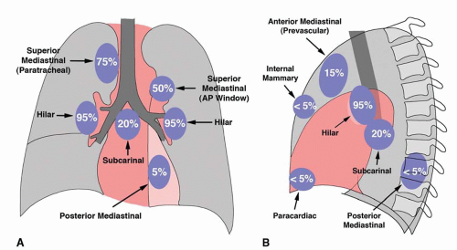
Chapter 8). The frequency of visible involvement of specific node groups is somewhat different on CT, as compared to plain radiographs. Specifically, hilar lymph node enlargement is not invariably seen on CT in patients with mediastinal adenopathy. In patients with enlarged nodes, CT shows abnormalities, in order of decreasing frequency, in the right paratracheal space (100%), aortopulmonary window (95%), hila (90%), subcarinal space (65%), prevascular space (50%), and posterior mediastinum (15%). Symmetry, or bilateral node involvement, is visible more often on CT than on radiographs.

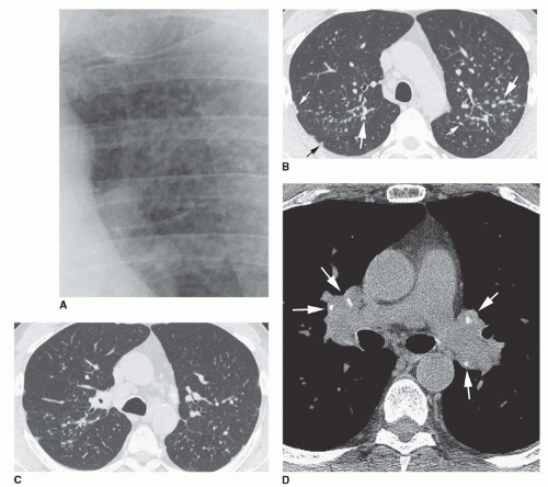
Lymph node masses in sarcoidosis may be quite large. Lymph node calcification is visible on CT in 25% to 50% of cases and may be hazy or dense or show a stippled or eggshell

appearance (Fig. 15-3D; see also Figs. 8-38A and 8-45 in Chapter 8). Rarely nodes appear necrotic or low in attenuation or enhance on CT.


FIG. 15.1. The 1-2-3 pattern of lymph node enlargement in sarcoidosis. A: On a chest radiograph, lymph node enlargement is visible in the right paratracheal mediastinum (1), right hilum (2), and left hilum (3). The presence of aortopulmonary window lymph node enlargement (apw) makes it a 1-2-3-4 pattern. B and C: On CT, lymph node enlargement is visible in the right paratracheal mediastinum (1), right hilum (2), and left hilum (3). Aortopulmonary window (apw) and subcarinal (subc) lymph node enlargement is also visible.


FIG. 15.2. Frequency of lymph node enlargement in patients with sarcoidosis as shown on posteroanterior (A) and lateral (B) radiographs.


FIG. 15.3. Pulmonary nodules in sarcoidosis. A: Detail view of the left upper lobe shows small, well-defined nodules. The lower lobes appeared normal. Left hilar enlargement is also seen. Most patients with lung nodules visible on radiographs also show lymph node enlargement. B: HRCT through the upper lobes shows a patchy perilymphatic distribution of sharply marginated nodules a few millimeters in diameter. The nodules involve the pleural surfaces and fissures (small arrows) and the peribronchovascular regions (large arrows). C: At a lower level, the nodules are less numerous, indicating an upper lobe predominance. D: Hilar lymph node enlargement with calcification (arrows) is visible using a soft-tissue window.


LUNG DISEASE


Nodules

In patients with sarcoidosis, pulmonary granulomas are found in relationship to lymphatics in the peribronchovascular interstitial space, subpleural interstitial space, and, to a lesser extent, the interlobular septa. This is termed a perilymphatic distribution (see Figs. 10-20 and 10-21 in Chapter 10). Although sarcoid granulomas are microscopic in size, they often coalesce to form macroscopic nodules several millimeters or more in diameter.


Plain Radiographs

Lung disease is visible on chest radiographs in about 40% of patients and is usually associated with lymph node enlargement (i.e., stage 2). Lung abnormalities are often bilateral and symmetrical as seen on radiographs, with an
upper lobe predominance in up to 80% of cases and a diffuse distribution in most others. A lower lobe predominance is unusual.

A nodular pattern is most common on chest radiographs, being seen in about half of the cases (see Figs. 15-3A and 15-6A). Nodules may appear well-defined or ill-defined and fuzzy and usually range from a few millimeters to 1 cm in diameter; a reticulonodular pattern is seen in an additional 25%.

Radiographs are relatively insensitive in showing lung abnormalities. In patients with radiographic stage 1 disease (i.e., no visible lung disease), 80% to 90% have lung lesions visible on high-resolution CT (HRCT) or on transbronchial biopsy.


HRCT

HRCT can show a variety of findings in patients with sarcoidosis (Table 15-1).

HRCT typically shows small nodules in patients with active disease. Nodules are as small as a few millimeters in diameter; they tend to be sharply defined despite their small size.


TABLE 15.1 HRCT Findings of Lung Disease in Sarcoidosis


Small sharply defined nodules


Typical of active lung disease


Perilymphatic distribution of nodules in most patients


Parahilar and peribronchovascular


Adjacent to fissures


Subpleural in the lung periphery


Interlobular septal nodules in some


Centrilobular peribronchovascular nodules in some


Patchy upper lobe predominance in most


Calcification may occur


Large nodules and masses


15%-25% of patients


Due to confluence of small granulomas


With or without air bronchograms


Commonly parahilar (peribronchvascular)


Associated with satellite nodules (i.e., the galaxy sign)


Ground-glass opacity


Uncommon


Due to numerous very small granulomas


Small nodules may be associated


Reticular opacities and fibrosis


Develops in 15% of patients


Peribronchovascular fibrosis with traction bronchiectasis


Upper lobe volume loss


Air-filled cysts


Honeycombing in some


Mycetoma may develop


Bronchial or bronchiolar abnormalities


Endobronchial granulomas


Atelectasis in some


Mosaic perfusion and air trapping

Only gold members can continue reading. Log In or Register to continue

Stay updated, free articles. Join our Telegram channel

Oct 10, 2016 | Posted by in CARDIOVASCULAR IMAGING | Comments Off on Sarcoidosis

Full access? Get Clinical Tree

Get Clinical Tree app for offline access