Fig. 28.1
Hepatic schistosomiasis. (a) Plain CT scanning demonstrates uneven low-density shadows in the left liver with poorly defined boundaries. (b, c) Contrast CT scanning demonstrates mildly uneven enhancement of the lesions in the arterial phase and portal vascular phase. (d) Postsurgical pathology demonstrates granuloma with accompanying deposition and calcification of schistosoma eggs
Case Study 2
A male patient aged 32 years complained of abdominal pain, aversion to cold, and fever for 2 weeks. He has a history of contact to contaminated water. His specific IgG antibody of schistosomiasis japonica was positive.
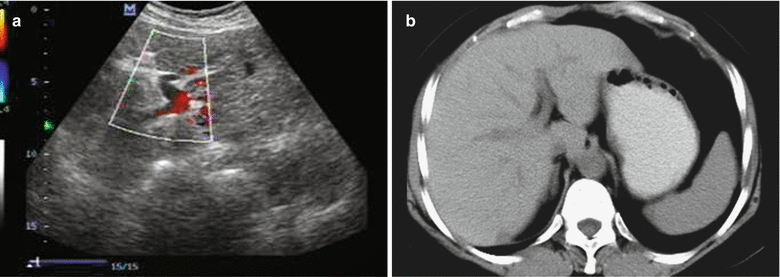

Fig. 28.2
Hepatic schistosomiasis. (a) Ultrasound demonstrates slightly increased echoes from the liver parenchyma, with thick and large light spots, and clear tubular network. (b) Plain CT scanning demonstrates slightly enlarged liver volume with even density
Case Study 3
A male patient aged 52 years had a medical history of schistosomiasis for more than 20 years.
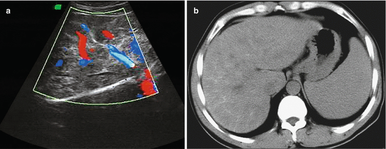

Fig. 28.3
Hepatic schistosomiasis. (a) Ultrasound demonstrates unsmooth Glisson’s capsule, thickened but uneven echoes from the liver parenchyma with maplike appearance, and poorly defined tubular network. Plain CT scanning demonstrates enlarged liver. (b) The liver is demonstrated with uneven density, multiple linear calcifications in the liver parenchyma with grid-like appearance, and splenomegaly
Case Study 4
A male patient aged 64 years had a medical history of schistosomiasis for 30 years.
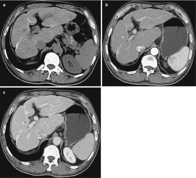

Fig. 28.4
Hepatic schistosomiasis. (a) Plain CT scanning demonstrates deformed liver with enlarged left and caudate lobes, increased density of the liver parenchyma, and strips of calcification at the liver and Glisson’s capsule. (b, c) Contrast CT scanning demonstrates even enhancement of the liver parenchyma and linear enhancement of intrahepatic septum
Case Study 5
A male patient aged 67 years had a medical history of schistosomiasis for more than 30 years. He complained of dull pain at the middle upper abdomen for more than 10 years.
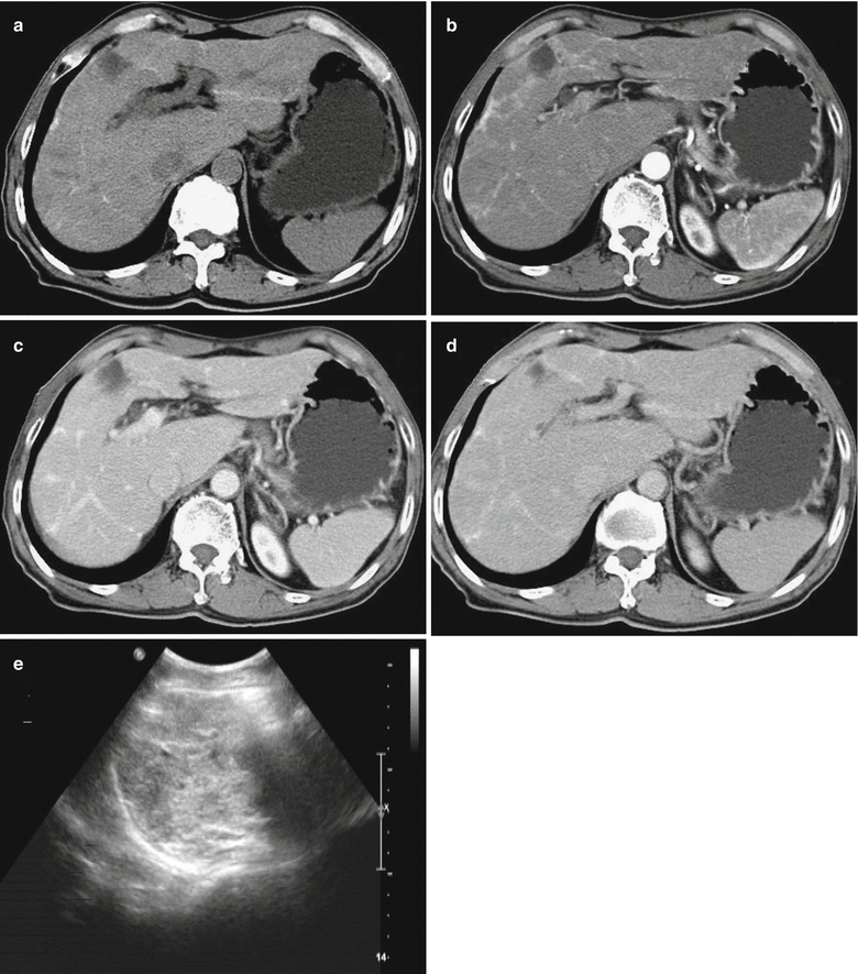

Fig. 28.5
Hepatic schistosomiasis. (a) Plain CT scanning demonstrates deformed liver and flakes of low-density lesions in the right liver lobe with maplike calcification. (b–d) Contrast CT scanning demonstrates no enhancement of the lesions in the right liver lobe and abnormal enhancement shadows of widened linear fibers in the liver parenchyma. (e) Ultrasound demonstrates thickened echoes from the liver parenchyma with maplike appearance
Case Study 6
A male patient aged 59 years had a medical history of schistosomiasis for more than 30 years.
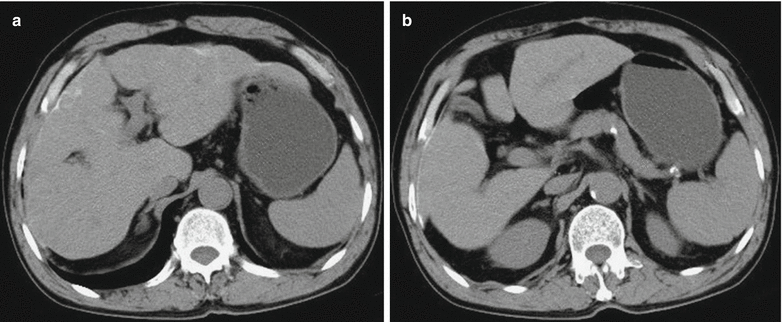

Fig. 28.6
Hepatic schistosomiasis. (a, b) Plain CT scanning demonstrates deformed liver, calcification of Glisson’s capsule, and mild splenomegaly
28.7.2 Pulmonary Schistosomiasis
28.7.2.1 X-Ray Radiology
Acute Pulmonary Schistosomiasis
Pulmonary lesions caused by acute schistosomiasis can be staged into early and late. The early pulmonary lesions are mechanical injuries caused by access of cercaria and adult worms into lung tissues and allergic responses of their metabolites. Most patients only experience increased and thickened lung markings and, in rare cases, small flakes and large flakes of shadows at bilateral middle and inferior lungs. Early lesions are characterized by early occurrence and rapid residing, which commonly last for about 2–3 weeks. They are subject to misdiagnosis by delayed examinations. Two to three months after the infection, the conditions develop into the late stage, with deposition of eggs in the pulmonary interstitium to form false nodules. At the late stage, miliary shadows with uneven density and different sizes may scatter in both lungs, which are poorly defined with a diameter of 2–5 mm. The lesions mostly distribute in the middle and lower lung fields, with some fusing into flakes. The lesions have a higher central density and a light peripheral density, resembling to alveolar edema. Some lesions may also fuse into snowflake-like lesion, with a diameter of about 7–8 mm.
Chronic Pulmonary Schistosomiasis
X-ray demonstrations are nonspecific, with mainly changes of pulmonary interstitium.
Changes of Pulmonary Interstitium
The demonstrations include blurry lung markings in bilateral middle and lower lungs, with spots of shadows or network of nodular shadows.
Pulmonary Infection
The lungs are demonstrated with large flakes of dense shadows with internal fluid level and poorly defined boundaries. The shadows can also be demonstrated in patches or cloud-like, with poorly defined boundaries. Intrapulmonary flakes of shadows can also be demonstrated with well-defined boundaries, resembling to those of inflammatory pseudoneoplasm.
Pulmonary Atelectasis
The lesion is commonly found at the fundus of lungs adjacent to the diaphragmatic surface, with strip-like or plate-like dense shadow in length of 2–5 cm and width of 1–2 cm. The lesion moves along with breathing, mostly occurring in patients of ascites type.
Pleural Effusion
The demonstrations include dull costophrenic angle, infrapulmonary effusion, or confined encapsulated effusion. Sometimes, pleural effusion is the only chest X-ray demonstration of chronic pulmonary schistosomiasis.
28.7.2.2 CT Scanning
In patients with acute pulmonary schistosomiasis, micronodular shadows can be transiently demonstrated and alveolar parenchyma is rarely found. In this stage, thickened bronchial wall at the lesions can also be found.
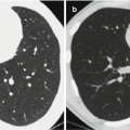
Chronic pulmonary schistosomiasis can be demonstrated by CT scanning with fissure-shaped exudation shadows in the lung fields, multiple fibrous stripes of shadows in the lungs, and typical nodular or micronodular shadows. The nodules are commonly distributed in the middle and lower lung fields, subpleural area, or bronchial bifurcations. The nodules have central high density with poorly defined boundaries and surrounding hyaloid exudation, in halo sign (Fig. 28.7). As the course of the disease prolongs, CT scanning can also demonstrate pulmonary interstitial fibrosis and pulmonary hypertension.
Case Study 7
A male patient aged 30 years complained of cough and chest pain for 2 days. By tests and examinations, the count of eosinophilic granulocytes in the peripheral blood significantly increased, and schistosome eggs were detected in the feces.
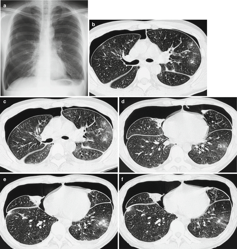

Fig. 28.7
Pulmonary schistosomiasis and pneumothorax. (a) X-ray demonstrates scattered patches of shadows in both lungs with poorly defined boundaries and bilateral pneumothorax. (b–f) Plain CT scanning demonstrates scattered nodular shadows in both lungs, with central high density and surrounding hyaloid changes, showing halo sign. There are also cord-like shadows that connect to the pleura and bilateral pneumothorax
28.7.3 Cerebral Schistosomiasis
Radiological demonstrations include multiple lesions in brain parenchyma, which are more common at the parenchyma of cerebral hemispheres, subcortex, and parietal lobe. The lesions are less commonly found at the brainstem, cerebellar hemispheres, cerebellopontine angle, and cerebral subdural space. In most cases, the main lesions are found at one certain area of the brain tissue. The lesions are multiple small nodules or nodules in different sizes that commonly fuse with each other. Contrast scanning demonstrates obvious even enhancement, with possible accompanying adjacent meningeal and vascular enhancements. In the lesions of schistosomiasis, sometimes calcification can be found in a typical target-like sign. In rare lesions, hemorrhage can be found.
28.7.3.1 CT Scanning
Plain CT scanning demonstrates low-density shadows of the lesions with surrounding low-density edema. Edema has a fingerstall-like distribution with significant space-occupying effect. By contrast scanning, the lesions are demonstrated with ring-shaped or mass-like enhancement. Dynamic contrast scanning demonstrates schistosomal granuloma with characteristic delayed enhancement, which is the most obvious during 5–15 min in delayed scanning. The characteristic demonstrations include slow enhancement and slow subsiding and fusion into masses. Perfusion CT scanning demonstrates significantly increased values of CBF, CBV, and PS of the schistosomal lesions, indicating significant vascular proliferation in the lesions, significant increased permeability, and severe damage of blood–cerebrospinal fluid barrier. In addition, MTT value is demonstrated with obvious decrease, indicating fast blood flow in the lesions, which is possibly related to the stimulation of inflammatory factors in the lesions as well as vascular dilations in the lesions.
28.7.3.2 MR Imaging
Plain MR imaging demonstrates equal or slightly low signal by T1WI and high or slightly high signal by T2WI, with obvious edema and space-occupying effect. Contrast imaging demonstrates various enhancements of the lesions, including nodular, sand-like, ring-like, or patchy enhancements, with multiple small nodules fusing into clusters. In the cases with enhancement at the cortex or subcortical interface between gray and white matters, there are surrounding large areas of edema, which extend toward the cortex-like fingerstall. DWI demonstrates the lesions with equal or slight high signals, which can be hardly distinguished from their surrounding edema and brain tissues. By ADC imaging, the lesions are demonstrated with increased ADC value, compared to the normal brain tissues. By eADC imaging, the lesions are demonstrated with decreased eADC value compared to the normal brain tissue.
28.7.3.3 Typing by CT and MRI
According to a series of pathological changes of cerebral schistosomiasis, some scholars have attempted to categorize the CT and MRI demonstrations of cerebral schistosomiasis. However, disagreement still exists. Peng RL et al. divided cerebral schistosomiasis into four types: encephalitis type, cerebral infarction type, granuloma type, and brain atrophy type. Sun JM et al. divided cerebral schistosomiasis into three types: encephalitis type, cerebral infarction type, and granuloma type, and they believed that localized brain atrophy is the common sequela of the above three types of cerebral schistosomiasis. We describe the detailed radiological demonstrations of each type as the following.
Encephalitis Type
This type is manifested as extensive edema of brain tissue induced by allergic responses and severe encephalitis due to toxins and metabolites secreted by schistosome eggs. No deposition of eggs can be found in the brain tissue. Plain CT plain demonstrates the lesions as low-density shadows with poorly defined boundaries. By MR imaging, the lesions are demonstrated as long T1 and long T2 signals. The lesions often involve multiple cerebral lobes and can be singular or multiple, which are more common at the parietal lobe. Contrast imaging demonstrates uneven patches of enhancement in the lesions. In the cases with pia mater involved, linear enhancement of meninges can be demonstrated (Figs. 28.8 and 28.9).
Granuloma Type
This type is the most common (Figs. 28.10 and 28.11). Based on the radiological demonstrations and the pathological findings of granuloma in the cases of cerebral schistosomiasis, Dong JN et al. further divided this type into four subtypes.
1.
Multiple small nodules type
The nodules have a diameter of 0.2–0.9 cm and have a scattering distribution or clusters of distribution, with no fusion of the nodules. By CT scanning and MR imaging, the cases of this type are demonstrated with stars-in-the-sky sign. The pathological basis of this type is acute nodules formed by worm eggs or small chronic nodules formed by worm eggs.
2.
Singular large nodule type
The singular nodule has a diameter of above 1.5 cm, with equal or slightly high density by plain CT scanning. The nodule clings to the gyrus, with a demonstration of gyrus hypertrophy. MR imaging demonstrates the nodule with equal or slightly low signal by T1WI and slightly high signal by T2WI. Contrast imaging demonstrates obvious uneven enhancement. Thin slice imaging demonstrates fusion of multiple small nodules with obvious enhancement into mass, which is the characteristic radiological demonstration of this type.
3.
Mixed nodule type
Both large nodules with a diameter of 1–3 cm and small nodules with a diameter of 0.2–0.9 cm are demonstrated with a scattering distribution. CT scanning and MR imaging demonstrate large nodules with enhancement with surrounding multiple small nodules.
4.


Nodule with ring-shaped enhancement type
Stay updated, free articles. Join our Telegram channel

Full access? Get Clinical Tree


