SECTION XIII VASCULAR IMAGING
Essentials 1
Case
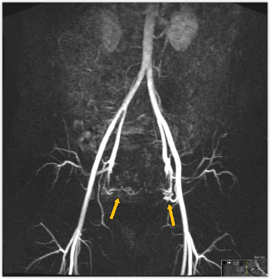
A 35-year-old woman with multiple uterine fibroids presents for pre-embolization workup. Shown here is the maximum intensity projection magnetic resonance angiography (MRA) image of the pelvis.
Questions
1. What vessels are demonstrated (arrows)?
A. Uterine branch of the ovarian artery
B. Helicine branches of the uterine artery
C. Inferior gluteal artery
D. Vaginal artery
E. Internal pudendal artery
2. Magnetic resonance imaging with magnetic resonance angiography is a time-consuming and expensive examination for evaluation of uterine leiomyomas. What are the specific indications for this study? (Select ALL that apply.)
A. Localization and characterization of fibroids
B. Determination of uterine vascular (arterial) anatomy
C. Measurement of vascular dimensions
D. Assessment of effectiveness of treatment (embolization)
E. Evaluation for presence of an arteriovenous shunt
3. True or False. Magnetic resonance angiography cannot be performed without gadolinium administration.
Answers and Explanations
Question 1
B. Correct! The arrows point to the helicine branches of the uterine artery. The uterine artery is a branch of the anterior division of the internal iliac artery. It courses lateral to medial in the base of the broad ligament and ascends along the lateral aspect of the body of the uterus. Helicine arteries (supply the uterus), ovarian branch (anastomoses with the ovarian artery), vaginal branch (anastomoses with the vaginal artery), and tubal branch (supplies the fallopian tubes) are the branches of the uterine artery. Helicine branches, which supply the uterus, have a characteristic corkscrew configuration, and this helps in identification.
Other choices and discussion
A. The ovarian artery arises anterolaterally from the abdominal aorta immediately below the level of the renal arteries. It courses in the retroperitoneum with the gonadal vein and ureter. In the pelvis, it courses through the ovarian suspensory ligament to supply the ovary. It anastomoses with the ovarian branch of the uterine artery but does not have a uterine branch.
C. The inferior gluteal artery is also a branch of the anterior division of the internal iliac artery. It traverses in the lateral pelvis and exits through the greater sciatic foramen to supply the gluteal region and thigh.
D. The vaginal artery arises from the anterior division of the internal iliac artery. It anastomoses with the vaginal branch of the uterine artery.
E. The internal pudendal artery is a branch of the anterior division of the internal iliac artery and arises anterior to the inferior gluteal artery. It exits the pelvis through the greater sciatic foramen and re-enters through the lesser sciatic foramen to supply the perineum.
Question 2
A. Correct! Although pelvic ultrasound is the initial imaging study for the evaluation of fibroids, magnetic resonance imaging (MRI) adds value, especially in cases of multiple fibroids for better demonstration of features such as size, location, and presence or absence of degeneration.
B. Correct! Determination of uterine vascular (arterial) anatomy is an essential step prior to embolization. Presence of an ovarian arterial supply results in unsuccessful embolization.
D. Correct! MRI can be performed after embolization to assess the degree of fibroid degeneration/necrosis and, therefore, the ultimate effectiveness of treatment.
E. Correct! Evaluating for the presence of an arteriovenous shunt is indispensable before embolization. There is a risk of shunting of the beads into the pulmonary arterial system in the presence of an arteriovenous shunt.
The other choice is incorrect. Measurement of the dimensions of the vessels is not required.
Question 3
False. Magnetic resonance angiography (MRA) can be performed even without the administration of intravenous contrast. This is extremely helpful in nephrotoxic patients, pregnant women, and in patients with contrast allergy. The three techniques to perform MRA without contrast include time-of-flight angiography, phase contrast imaging, and three-dimensional electrocardiograph-triggered half Fourier fast spin echo. Generally, these techniques take longer to acquire compared to postcontrast angiography.
Suggested Readings
Bulman JC, Ascher SM, Spies JB. Current concepts in uterine fibroid embolization. Radiographics 2012;32(6):1735–1750 Stepansky F, Hecht EM, Rivera R, et al. Dynamic MR angiography of upper extremity vascular disease: pictorial review. Radiographics 2008;28:e28Top Tips
Helicine branches of the uterine artery provide blood supply to the uterus. They have a typical corkscrew appearance.
MRI and MRA of the pelvis is the imaging tool of choice for potential uterine fibroid embolization candidates. MRI performed before and after uterine fibroid embolization is critical for demonstrating and localizing leiomyomas, identifying red flags regarding vascular supply, assessing the likelihood of symptom relief after therapy on the basis of imaging characteristics, and monitoring the response of leiomyomas to therapy.
MRA can be performed without the use of intravenous contrast.
Essentials 2
Case
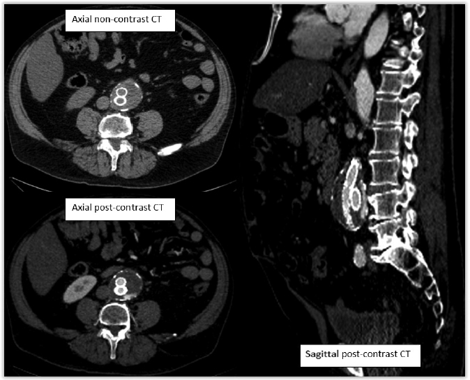
A 64-year-old man had a follow up computed tomography angiography 6 months after aortic stent graft placement for aneurysm.
Questions
1. What is the diagnosis?
A. Type I endoleak
B. Type II endoleak
C. Type III endoleak
D. Type IV endoleak
E. Type V endoleak
2. What is the overall most common type of endoleak?
A. Type I endoleak
B. Type II endoleak
C. Type III endoleak
D. Type IV endoleak
E. Type V endoleak
3. Which type(s) of endoleak requires immediate intervention? (Select ALL that apply.)
A. Type I endoleak
B. Type II endoleak
C. Type III endoleak
D. Type IV endoleak
E. Type V endoleak
Answers and Explanations
Question 1
B. Correct! Type II endoleak occurs due to retrograde flow of blood into the aneurysmal sac from branches (inferior mesenteric artery, lumbar arteries) of the aorta. It is most commonly encountered in abdominal aortic aneurysms. Contrast located in the periphery of the aneurysmal sac without communication with the stent is indicative of a type II endoleak. If located anteriorly, the inferior mesenteric artery is the culprit, whereas those located posterolaterally arise from the lumbar arteries. Another diagnostic clue is tubular configuration of the contrast abutting the aortic wall on coronal or sagittal reformatted images.
Other choices and discussion
A. Type I endoleak occurs due to inadequate apposition between the stent graft and the aneurysm sac. On computed tomography angiography, the diagnosis of type I endoleak is made when contrast is identified in the aneurysmal sac in communication with the proximal or distal attachment sites.
C. Type III endoleak is due to defective components of the endograft, such as stent graft fractures or rupture/tear of the graft material. The underlying cause is proposed to be repetitive stress on the graft from arterial pulsations. Contrast pooling around the stent graft with peripheral sparing is suggestive of type III endoleak.
D. Type IV endoleak is a diagnosis of exclusion and is due to stent-graft porosity. These occur at the time of implantation and are self-limited once the coagulation profile is normalized.
E. Type V endoleak, also called endotension, is defined as an increase in size of the aneurysmal sac without imaging evidence of endoleak.
Question 2
B. Correct! Type II endoleak is the overall most common type of endoleak encountered in clinical practice (40%). Of note, type I endoleak is the most common type of endoleak that occurs in thoracic aortic aneurysms.
The other choices are incorrect.
Question 3
A. Correct! Type I endoleak represents direct communication between the systemic arterial system and the aneurysmal sac, and is therefore at greater risk of rupture of the sac. They are usually treated by securing the attachment sites with balloons, stents, or stent graft extensions.
C. Correct! Type III endoleak also represents direct communication between the high-pressure arteries and the native aneurysmal sac and therefore requires immediate intervention. The defect is usually covered with stent graft extension.
Other choices and discussion
B. A total of 40% of type II endoleaks spontaneously thrombose. They are repaired if the patient is symptomatic or if the sac increases in size over time.
D. Type IV endoleak occurs at the time of implantation and autocorrects after normalization of the coagulation profile. Therefore, no treatment is necessary.
E. Once endotension (type V endoleak) is confirmed, open surgical repair is required. However, it is not an emergency.
Suggested Readings
Bashire MA, Ferral H, Jacobs C, et al. Endoleaks after endovascular abdominal aortic aneurysm repair: management strategies according to CT findings. Am J Roentgenol 2009;193:W178–186 Stavropoulos SW, Charagundla SR. Imaging techniques for detection and management of endoleaks after endovascular aortic aneurysm repair. Radiographics 2007;243:641–655Top Tips
Type II endoleak is the most common type of endoleak.
Type I and III endoleaks require emergent treatment.
Type IV endoleak is self-limited and does not require treatment. Type II and V leaks can be managed conservatively with close follow up. Treatment is necessary if the patient has symptoms or if the aneurysmal sac continues to increase in size.
Essentials 3
Case
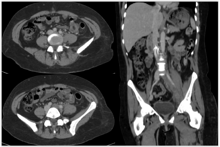
A 45-year-old woman presents with a 5-day history of progressive left lower extremity swelling. Computed tomography of the abdomen and pelvis with contrast was performed. Venous phase images are shown.
Questions
1. Where is the abnormality?
A. Inferior vena cava
B. Left common iliac artery
C. Left common iliac vein
D. Right common iliac vein
2. What is the diagnosis?
A. Paget-Schroetter syndrome
B. Lemierre syndrome
C. May-Thurner syndrome
D. Budd-Chiari syndrome
3. What is the appropriate management of May-Thurner syndrome?
A. Systemic anticoagulation
B. Catheter-directed thrombolysis and endovascular stent placement
C. Catheter-directed thrombolysis alone
D. Nothing
Answers and Explanations
Question 1
C. Correct! There is a large occlusive thrombus in the left common iliac vein.
The other choices are incorrect.
Question 2
C. Correct! May-Thurner syndrome is alternatively called Cockett syndrome. It is characterized by compression of the left common iliac vein by the right common iliac artery against the vertebral body. This in turn results in left lower extremity swelling with or without thrombosis of the left common iliac vein. It generally affects young and middle-aged adults and predominantly women. The presence of a pelvic mass must be excluded as the cause of thrombosis before making this diagnosis.
Other choices and discussion
A. Paget-Schroetter syndrome is a thoracic outlet syndrome characterized by subclavian vein thrombosis between the first rib and the clavicle. It is also referred to as effort thrombosis and is typically seen in young athletic males. It is thought to be due to vigorous overhead arm physical activity.
B. Lemierre syndrome, also called postanginal sepsis, is characterized by thrombophlebitis of the internal jugular vein secondary to adjacent oropharyngeal infection. The critical feature is the spread of infection into the chest that manifests as pulmonary septic emboli.
D. The key imaging findings in Budd-Chiari syndrome are thrombosis of the hepatic veins and/or inferior vena cava.
Question 3
B. Correct! Catheter-directed thrombolysis followed by endovascular stent placement has evolved as an alternate to surgery and is now the preferred management technique. It has a high success rate of 95% and excellent 1-year patency rates of 90 to 100% (mean 96%).
Other choices and discussion
A. Systemic anticoagulation was the first line of treatment prior to thrombolysis. It is no longer used due to a very low patency rate. Oral anticoagulants are used after stent placement to prevent recurrence of thrombosis.
C. Catheter-directed thrombolysis alone is effective and has a higher patency rate at 6 months than oral anticoagulation alone. However, a recurrence rate of 73% has been reported when the primary cause of obstruction has not been addressed.
D. May-Thurner syndrome can lead to dreadful complications such as pulmonary embolism and acute limb ischemia. Therefore, treatment is necessary.
Suggested Readings
Eliahou R, Sosna J, Bloom AI. Between a rock and a hard place: clinical and imaging features of vascular compression syndromes. Radiographics 2012;32:E33–49 Lamba R, Tanner DT, Sekhon S, et al. Multidetector CT of vascular compression syndromes in the abdomen and pelvis. Radiographics 2014;34:93–115Top Tips
Compression with or without thrombosis of the left common iliac vein by the right common iliac artery is defined as May-Thurner syndrome.
Catheter-directed thrombolysis with endovascular stent placement has emerged as the treatment of choice.
Pulmonary embolism and acute limb ischemia (phlegmasia cerulea dolens) are the two potential complications.
Essentials 4
Case
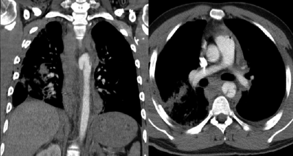
A 35-year-old man is status post a high-speed motor vehicle accident. Images are from chest computed tomography with contrast.
Questions
1. What is the diagnosis?
A. Aortic dissection
B. Intimal tear
C. Normal ductus diverticulum
D. Aortic transection with pseudoaneurysm
2. What is the MOST common location for this entity?
A. Diaphragmatic hiatus
B. Aortic root
C. Aortic isthmus
D. Ascending aorta
3. What is the treatment for aortic transection with pseudoaneurysm?
A. Observation
B. Open surgical repair
C. Angioplasty
D. Endovascular stent graft placement
Answers and Explanations
Question 1
D. Correct! This is a classic case of aortic transaction with pseudoaneurysm formation. The direct signs of abnormal contour and outpouching of the aorta representing the pseudoaneurysm are noted. In addition, the indirect sign of the presence of periaortic hematoma without a fat plane between the hematoma and the aorta is also seen.
Other choices and discussion
A. Aortic dissection is an extremely rare sequelae of trauma. It is characterized by intimal tear with resultant accumulation of blood between the intima and the adventitia, and the formation of a second false lumen. On imaging, an intimal flap and two lumens are the clues for diagnosis.
B. Intimal tear in the trauma setting is categorized under minimal aortic injury (MAI). It is characterized by intimal flap or intraluminal aortic thrombus. Intramural hematoma also falls under the same category. The diagnosis of MAI has increased since the advent of thin-slice computed tomography.
C. Although this is the typical location for a ductus diverticulum, the presence of periaortic hematoma precludes that diagnosis. Other clues of a ductus diverticulum include a smooth contour and obtuse margins of the diverticulum with the aortic wall.
Question 2
C. Correct! Aortic transection occurs more commonly (90%) at the isthmus because of the relative immobility of this region due to tethering by the ligamentum arteriosum. This results in shearing injury during high-speed motor vehicle collisions.
Other choices and discussion
A. A total of 5% of acute traumatic aortic injuries occur in the region of the diaphragmatic hiatus.
B. The aortic root is not a typical location for an acute traumatic injury.
D. A total of 5% of acute traumatic aortic injuries occur in the ascending aorta.
Question 3
D. Correct! Endovascular stent graft placement has recently emerged as the treatment of choice. The advantages of this method over open repair include the avoidance of single lung ventilation, aortic cross-clamping, cardiopulmonary bypass, and systemic anticoagulation. In addition, with endovascular stent graft placement, there is reduced blood loss and reduced surgical time.
Other choices and discussion
A. Conservative management is reserved for patients with MAI or patients with other critical comorbid injuries.
B. Open surgical repair has been the treatment of choice for decades. However, its use is fading due to the associated risks of anesthesia and the surgery itself.
C. Angioplasty is not an appropriate treatment for aortic injury.
Suggested Readings
Cullen EL, Lantz EJ, Johnson M, et al. Traumatic aortic injury: CT findings, mimics, and therapeutic options. Cardiovasc Diagn Ther 2014;4:238–244 Morgan TA, Steenburg SD, Siegel EL, et al. Acute traumatic aortic injuries: posttherapy multidetector CT findings. Radiographics 2010;44:158–163 Steenburg SD, Ravenel JG, Ikonomidis JS, et al. Acute traumatic aortic injury: imaging evaluation and management. Radiology 2008;248:748–762Top Tips
Acute traumatic aortic injury occurs most commonly at the aortic isthmus.
Endovascular therapy is now the treatment of choice.
Pretherapy multidetector computed tomography should document the following: caliber of the aorta proximal and distal to the injury, distance from the left subclavian artery to the injury, length of the vascular injury, and presence of any anatomic variants.
Essentials 5
Case
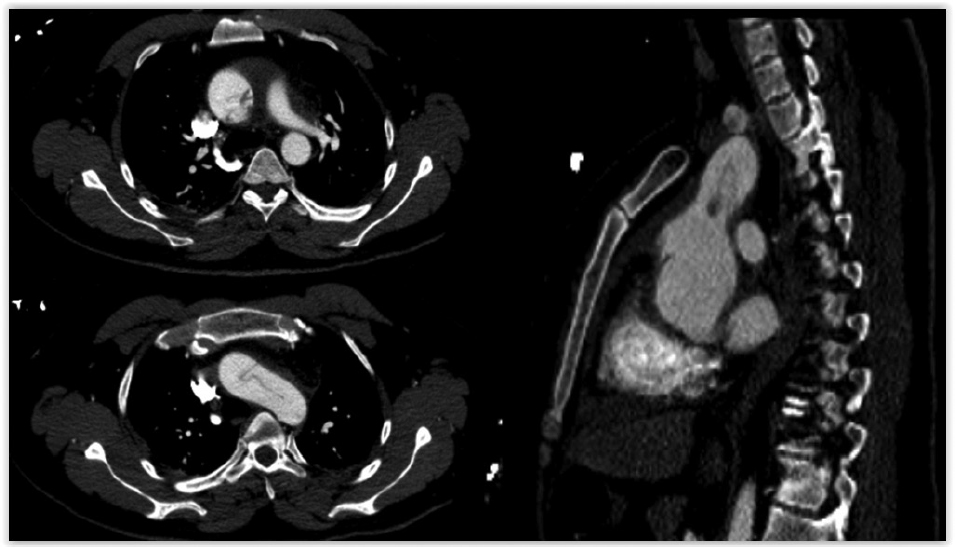
A 61-year-old man presents to the emergency room with severe acute chest pain. Computed tomography of the chest was performed.
Questions
1. What is the diagnosis?
A. Stanford type A dissection
B. Stanford type B dissection
C. DeBakey type III dissection
D. Stanford type C dissection
2. True or False. All dissections of the type presented in this case are typically treated surgically.
3. What are the primary indications for repair of type B dissection? (Select ALL that apply.)
A. All type B dissections
B. Ruptured aorta or descending aortic diameter > 6 cm
C. Renal or visceral vascular compromise
D. Uncontrolled hypertension
Answers and Explanations
Question 1
A. Correct! Two classifications exist for aortic dissection: Stanford and DeBakey. Aortic dissections involving the ascending aorta regardless of the distal extent are referred to as Stanford type A dissections. Sixty percent of dissections are type A. Acute aortic dissections are those diagnosed within 14 days of the onset of symptoms, whereas chronic dissections are older than 14 days. Hypertension is the most common risk factor for aortic dissection. Other contributing factors include pregnancy, aortic stenosis, or the presence of connective tissue disorders such as Marfan syndrome, cystic medial necrosis, Ehlers-Danlos syndrome, and Turner syndrome.
Other choices and discussion
B. Aortic dissection distal to the origin of the left subclavian artery with sparing of the ascending aorta is termed a Stanford type B dissection.
C. DeBakey type III dissection also involves the descending thoracic aorta distal to the origin of the left subclavian artery (this is similar to a Stanford type B). DeBakey type I involves both ascending and descending aorta, whereas type II involves only the ascending aorta (both come under Stanford type A).
D. Stanford type C dissection does not exist.
Question 2
True. All type A dissections require immediate intervention. The dreaded complications of type A dissection include rupture of the dissection into the pericardium with progressive pericardial tamponade, occlusion of the supra-aortic vessels, extension of the dissection into the coronary arteries resulting in ischemia, and severe aortic insufficiency with acute heart failure.
Question 3
B. Correct! Irregularity of the aortic wall, extravasation of contrast, hemothorax, hemopericardium, and hyperattenuating mediastinal fluid collections are indicative of aortic rupture, when immediate intervention is required. A descending aortic diameter of > 6 cm is at great risk for rupture, and therefore, necessitates emergent intervention.
C. Correct! Extension of the intimal flap into the branches of the aorta (renal, celiac, superior and inferior mesenteric arteries) can cause end-organ ischemia. This is considered a surgical emergency.
D. Correct! Most type B dissections are managed conservatively by controlling the blood pressure. However, in patients with hypertension refractory to medical therapy, surgical management is necessary to prevent rupture.
The other choice is incorrect. Approximately 40% of the dissections are type B in nature. Most of these can be treated conservatively.
Suggested Reading
Sebastia C, Pallisa E, Quiroga S, et al. Aortic Dissection: diagnosis and follow-up with helical CT. Radiographics 1999;19:45–60Top Tips
Aortic dissections have two classifications: Stanford and DeBakey. Stanford is the most commonly used classification.
Stanford type A dissections require immediate intervention.
Stanford type B dissections are treated conservatively, unless there is uncontrolled hypertension, end-organ ischemia, ruptured aorta, or aortic diameter > 6 cm.
Essentials 6
Case
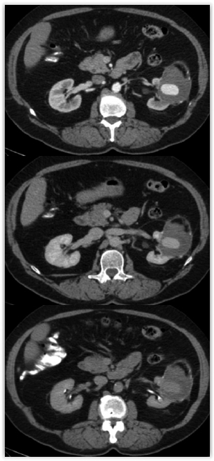
A 66-year-old man presents for evaluation of left flank pain. He is status-post renal biopsy one day earlier. Computed tomography of the abdomen and pelvis with contrast was performed.
Questions
1. What is the diagnosis?
A. Active extravasation of contrast
B. Subcapsular hematoma
C. Pseudoaneurysm
D. True aneurysm
2. What is the MOST common nontraumatic abdominal visceral arterial aneurysm?
A. Hepatic artery
B. Celiac artery
C. Superior mesenteric artery
D. Splenic artery
3. Which of these visceral arterial aneurysms requires intervention regardless of size in otherwise normal patients?
A. Hepatic artery
B. Renal artery
C. Superior mesenteric artery
D. Splenic artery
Answers and Explanations
Question 1
C. Correct! This patient has a pseudoaneurysm. Pseudoaneurysm is an entity seen secondary to trauma, infection, or iatrogenic causes such as percutaneous biopsy or drainage. It is the result of partial or complete disruption of the aortic wall with resultant formation of a sac contained by media, adventitia, or connective tissue, or simply by soft tissue structures surrounding the vessel wall. On computed tomography, it appears as a smooth, well-circumscribed round or oval-shaped focus of contrast that does not enlarge or increase in attenuation on the more delayed images. Pseudoaneurysms have a greater risk of rupture due to the deficient arterial wall. Therefore, immediate intervention is warranted.
Other choices and discussion
A. Active extravasation of contrast is depicted as an irregularly shaped pooling of contrast with ill-defined edges on the arterial phase that increases in size on the venous and delayed phases. An adjacent hematoma with various stages of blood products is usually associated with active extravasation. Treatment includes angiographic embolization.
B. Subcapsular hematoma is a well-known complication of renal biopsy. It is represented as a curvilinear high-density hematoma surrounding the kidney. Because of the subcapsular location of the hematoma, there is compression of the renal parenchyma, which can result in a Page kidney. Page kidney is defined as the phenomenon of developing hypertension due to compression of the renal parenchyma and vasculature with resultant activation of the renin-angiotensin system.
D. True aneurysms are typically seen with atherosclerotic disease. They have all three layers of the vessel wall intact. They are usually fusiform in shape, whereas pseudoaneurysms are saccular. Visceral arterial aneurysms are less common than visceral pseudoaneurysms, especially with a history of trauma or surgical intervention.
Question 2
D. Correct! Splenic artery aneurysm is the most common type of abdominal visceral arterial aneurysm and accounts for 60 to 80% of cases. It has a male-to-female ratio of 1:4. Predisposing conditions include pregnancy, multiparity, portal hypertension, systemic hypertension, medial fibroplasia, and alpha-1 antitrypsin deficiency. Rupture is seen in 3 to 10% of cases. The mortality rate in nonpregnant patients is 10 to 25%, but that rate increases to approximately 70% during pregnancy.
Other choices and discussion
A. True aneurysms involving the common hepatic artery, hepatic artery proper, or intrahepatic arterial branches account for 20% of the cases. They are the second most common type of abdominal visceral arterial true aneurysm.
B. Celiac artery aneurysm is the fourth most common type of visceral arterial true aneurysm. They can be associated with abdominal aortic aneurysms in up to 18% of cases and with other visceral arterial aneurysms in about 50% cases. Therefore, presence of one aneurysm necessitates a search for another.
C. Superior mesenteric artery aneurysm is the third most common type of nontraumatic visceral arterial aneurysm. Unlike other visceral artery aneurysms, 70 to 90% of these aneurysms are symptomatic and associated with a high incidence of ischemic bowel complications.
Question 3
C. Correct! Unlike other visceral artery aneurysms, 70 to 90% of superior mesenteric artery aneurysms are symptomatic and associated with a high incidence of ischemic bowel complications. The risk of rupture is estimated to be 50%, and the surgical mortality rate in the setting of a ruptured aneurysm is 38%. On the other hand, no deaths have been reported in cases with elective intervention. The success rates for treatment of these aneurysms vary from 75 to 100%. Considering all of these factors, superior mesenteric artery aneurysms are treated irrespective of the size.
Other choices and discussion
A. Hepatic arterial true aneurysms are treated if the patient is symptomatic or when the size exceeds 2 cm. However, in patients with polyarteritis nodosa or fibromuscular dysplasia, treatment is recommended regardless of the size. The mortality rate associated with rupture is 21%.
B. Intervention is required in renal arterial aneurysms if the size is > 2 cm or if the patient is symptomatic. However, treatment is deemed necessary in pregnant women regardless of the size.
D. Symptomatic patients and splenic artery aneurysms > 2 cm require intervention in otherwise normal patients. However, with pregnancy, portal hypertension, connective tissue disorders, and alpha-1 antitrypsin deficiency, treatment is initiated regardless of the size.
Suggested Readings
Jesinger RA, Thoreson AA Lamba R, et al. Abdominal and pelvic aneurysms and pseudoaneurysms: imaging review with clinical, radiologic, and treatment correlation. Radiographics 2013;33:E71–96 Saad EA, Saad WEA, Davies MG, et al. Pseudoaneurysms and the role of minimally invasive techniques in their management. Radiographics 2005;25(Suppl 1):S173–189Top Tips
Pseudoaneurysms are more common than true aneurysms in the setting of trauma, infection, or iatrogenic etiologies. Pseudoaneurysms require emergent intervention due to high risk of rupture.
Splenic artery aneurysm is the most common type of visceral arterial true aneurysm.
All superior mesenteric arterial true aneurysms require elective intervention.
Essentials 7
Case
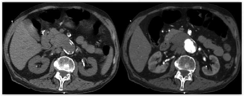
A 77-year-old man with chronic history of abdominal pain had a computed tomography angiogram of the abdomen and pelvis with contrast performed. Pre- and postcontrast axial computed tomography images are shown.
Questions
1. What is the most complete diagnosis?
A. Abdominal aortic aneurysm
B. Abdominal aortic aneurysm with impending rupture
C. Abdominal aortic aneurysm with contained rupture
D. Aortoenteric fistula
2. At what size are abdominal aortic aneurysms typically treated?
A. 3 cm
B. 4 cm
C. 5 cm
D. 6 cm
3. What is the MOST common complication after endovascular stent-graft placement for abdominal aortic aneurysm?
A. Endoleak
B. Graft thrombosis
C. Shower embolism
D. Graft infection
Answers and Explanations
Question 1
C. Correct! This patient has an abdominal aortic aneurysm with a contained rupture.
The computed tomography (CT) features of chronic contained rupture include discontinuity of the wall or rim calcification of the aneurysm, well-defined soft tissue density adjacent to the aorta (as seen here), displaced viscera, and no contrast material in the hematoma. Draping of the posterior aspect of the aorta over the adjacent vertebral body is suggestive of a contained rupture of the posterior wall of the aorta and is termed the “draped aorta” sign. Frank and acute ruptures demonstrate significant retroperitoneal hemorrhage with or without contrast extravasation.
Other choices and discussion
A. There is abnormal dilatation of the abdominal aorta, consistent with an aneurysm. However, there is more here than just a simple aneurysm.
B. A well-defined peripheral crescent of increased attenuation within the thrombus of a large abdominal aortic aneurysm on a noncontrast study is a CT sign of acute or impending rupture. It is the earliest and most specific imaging manifestation of impending rupture, but is not seen here.
D. In the presence of a patent fistula, CT with contrast would demonstrate extravasation of contrast into the adjacent bowel. In this case, the duodenum is displaced, but there is no contrast material in the lumen.
Question 2
C. Correct! Abdominal aortic aneurysms ≥ 5 cm in size are associated with a greater risk of rupture and thus are often treated at this size. The other criterion is > 10 mm increase in size of the aneurysm per year. With this criterion, aneurysms < 5 cm are also treated.
The other choices are incorrect.
Question 3
A. Correct! Endoleak is the most common complication. Leakage into the aneurysm status post stent graft placement is referred to as an endoleak. This is the most common complication after endovascular repair of abdominal aortic aneurysm, with rates ranging from 2.4 to 45.5%. Five types of endoleak have been described. Types I and III require immediate intervention. Type II endoleak is the most common type of endoleak.
Other choices and discussion
B. The rate of graft thrombosis ranges from 3 to 19%. The thrombosis can be parietal, circular, or semicircular within the stent-graft. Short-term follow up studies are performed to determine the prognosis.
C. Shower embolism is one of the most serious complications of abdominal aortic aneurysm repair and is seen more commonly with endovascular repair than with conventional surgery. This condition is associated with high perioperative mortality.
D. Graft infection with pseudoaneurysm formation is also a rare complication associated with high morbidity and mortality rates.
Suggested Readings
Mita T, Arita T, Matsunaga N, et al. Complications of endovascular repair for thoracic and abdominal aortic aneurysm: an imaging spectrum. Radiographics 2000;20:1263–1278 Rakita D, Newatia A, Hines JJ, et al. Spectrum of CT findings in rupture and impending rupture of abdominal aortic aneurysms. Radiographics 2007;27:497–507Top Tips
A high attenuation crescent sign is indicative of impending rupture of an abdominal aortic aneurysm.
The draped aorta sign is suggestive of a contained rupture of an aortic aneurysm.
Endoleak is the most common complication after abdominal aortic aneurysm repair.
Essentials 8
Case
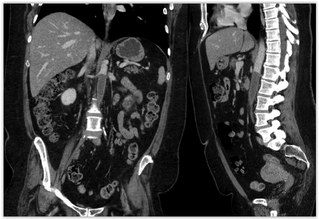
A 45-year-old man had a computed tomographic angiography performed. Coronal and sagittal reformatted images are shown.
Questions
1. What is the classic clinical triad of symptoms that patients with this diagnosis present with?
A. Paraplegia, pain, and paresthesia
B. Thigh, hip, or buttock claudication, absent/diminished femoral pulse, and impotence
C. Thigh, hip, or buttock claudication, absent/diminished femoral pulse, and quadriplegia
D. Abdominal pain, back pain, and impotence
2. Which of the following best describes midaortic dysplastic syndrome?
A. Aneurysmal dilatation of the abdominal aorta and its branches
B. Thrombotic occlusion of the abdominal aorta and its branches
C. Narrowing of the abdominal aorta and its branches
D. Dissection of the abdominal aorta extending into its branches
3. What is the BEST diagnostic study for evaluation of abdominal aortic stenotic/occlusive disease?
A. Computed tomographic angiography
B. Ultrasound with Doppler
C. Magnetic resonance angiography
D. Digital subtraction angiography
Answers and Explanations
Question 1
B. Correct! These images demonstrate thrombotic occlusion of the infrarenal abdominal aorta, which is consistent with Leriche syndrome. The classic clinical triad of Leriche syndrome includes thigh, hip, or buttock claudication; absent/diminished femoral pulse; and impotence. Three main subtypes have been described based on the location of the thrombus: juxtarenal or within 5 mm of the lower renal arterial origin; infrarenal or cephalic to the origin of the inferior mesenteric artery; and inframesenteric, which is caudal to the origin of the inferior mesenteric artery. The etiology of Leriche syndrome is atherosclerotic disease.
The other choices are incorrect.
Question 2
C. Correct! Midaortic dysplastic syndrome (also called abdominal aortic coarctation) is characterized by narrowing of the abdominal aorta and its branches. It is an uncommon condition typically affecting children and young adults. Refractory hypertension and a weakened or absent femoral pulse are the presenting features of this entity. These patients die by age 35 to 40 due to progressive hypertension, if not treated. Aortic reconstruction with prosthetic or autologous venous grafts is a treatment option.
The other choices are incorrect.
Question 3
A. Correct! Computed tomographic angiography (CTA) is best diagnostic modality for evaluation of arterial (aortic) pathology because of the great spatial resolution, fast acquisition time (requiring less time), and ability to reconstruct and reformat the images. Radiation is the precluding factor for CTA in pregnant women and children. The contraindications for CTA include contrast allergy and renal failure.
Other choices and discussion
B. Ultrasound with Doppler is good for evaluation of the peripheral arteries and the superficial and deep venous structures. Deeper structures such as the aorta may be difficult to visualize due to overlying bowel gas. Therefore, ultrasound is not a reliable modality for accurate evaluation of the abdominal aorta.
C. Magnetic resonance angiography (MRA) has good contrast resolution but has lower spatial resolution than CTA. The major disadvantages of MRA include long acquisition time, resultant motion artifacts, and greater cost. Contraindications for MRA are contrast allergy, pregnancy, and renal failure. MRA without contrast (time-of-flight or phase contrast angiography) can be performed in nephrotoxic patients. Generally, scan time is longer with these techniques compared to postcontrast MRA.
D. Digital subtraction angiography is a gold standard for arterial pathology. However, it is invasive and is not generally used for diagnostic purposes since the advent of CTA.
Suggested Readings
Bhatti AM, Mansoor J, Younis U, Siddique K, Chatta S. Mid aortic syndrome: a rare vascular disorder. J Pak Med Assoc 2011;61:1018–1020 Sebastia C, Quiroga S, Boye R, et al. Aortic stenosis: spectrum of diseases depicted at multisection CT. Radiographics 2003;23:S79–S91Top Tips
Leriche syndrome is atherosclerotic occlusion of the infrarenal abdominal aorta with a classic clinical triad of thigh, hip, or buttock claudication; absent/diminished femoral pulse; and impotence.
Midaortic dysplastic syndrome (also called coarctation of the abdominal aorta) is characterized by narrowing of the abdominal aorta and its branches. Children and young adults present with uncontrolled hypertension.
CTA is the diagnostic modality of choice for abdominal aortic pathology.
Essentials 9
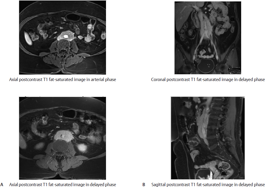
Questions
1. What is the diagnosis?
A. Retroperitoneal hematoma
B. Infectious aortitis
C. Perianeurysmal fibrosis
D. Retroperitoneal fibrosis
2. What is the classic feature suggestive of this disorder on intravenous pyelography and retrograde pyelography?
A. Lateral bowing of the ureters
B. Medial deviation of the ureters
C. Delayed excretion of contrast by the ureters
D. Hydronephrosis
3. True or False. A catheter can be easily passed through the narrowed area of the ureter in retroperitoneal fibrosis, unlike in the presence of intrinsic or extrinsic obstructive ureteral lesions.
Answers and Explanations
Question 1
D. Correct! This patient has retroperitoneal fibrosis (RPF; aka Ormond disease). RPF is characterized by proliferation of fibroinflammatory tissue typically surrounding the infrarenal abdominal aorta, inferior vena cava, and iliac vessels. It can extend to neighboring structures, especially the ureters, and ultimately lead to renal failure. Two subtypes of RPF have been described: idiopathic and secondary. Idiopathic RPF is an autoimmune condition and can lead to obstructive uropathy in 56 to 100% of patients, deep vein thrombosis (due to entrapment of retroperitoneal lymphatics), and varicocele (secondary to gonadal vein involvement) formation. The secondary form can be benign (from ingestion of drugs) or malignant. Malignant RPF is a described as a desmoplastic response to retroperitoneal primary tumors (lymphoma or sarcoma) or metastatic disease (breast, colon, stomach, lung, thyroid, carcinoid, and genitourinary tract).
Owing to the poor prognosis of malignant RPF, it is important to differentiate benign from malignant RPF. Anterior displacement of the aorta from the spine is usually seen with malignant RPF; however, there are exceptions (as in this case). In idiopathic RPF, the mass envelopes the retroperitoneal structures rather than exerting mass effect, as in malignant RPF. A retroperitoneal mass that has low signal intensity on T2-weighted images is highly suggestive of benign RPF in the late inactive stage. On the other hand, high T2 signal intensity is seen in both malignant and early stage idiopathic RPF, and this appearance is therefore nonspecific. Ultimately, histopathologic examination is often mandatory to establish the definitive diagnosis in suspicious cases with a possibility of malignancy. The mainstay of treatment for idiopathic RPF is steroid administration.
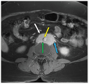
This postcontrast T1 fat-saturated image in delayed phase demonstrates an enhancing soft tissue mass (white arrow) encasing the infrarenal abdominal aorta (yellow arrow) and the left ureter (blue arrow). There is elevation of the posterior wall of the aorta from the vertebral body (green arrow), which is unsual for idiopathic retroperitoneal fibrosis.
Stay updated, free articles. Join our Telegram channel

Full access? Get Clinical Tree


