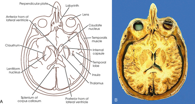
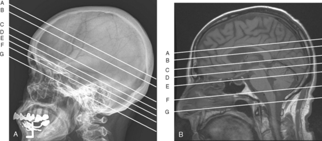
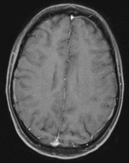
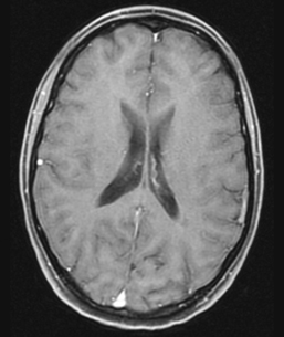
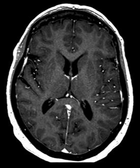
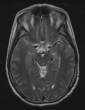
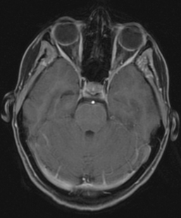
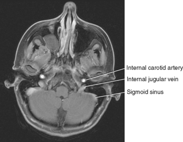
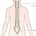
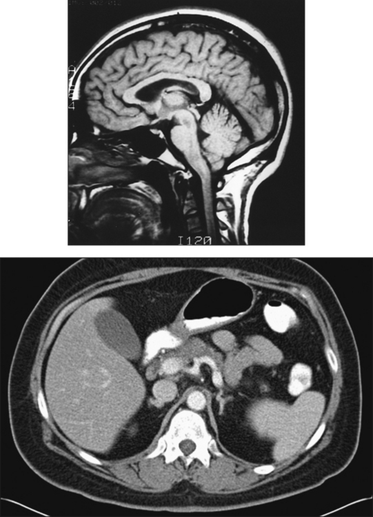
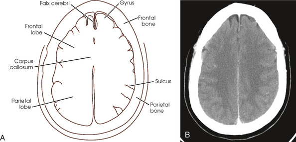
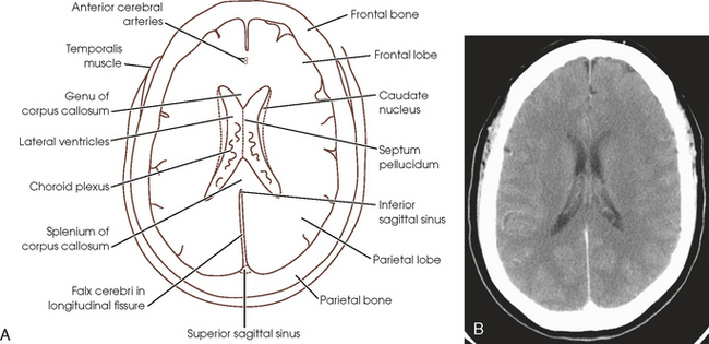
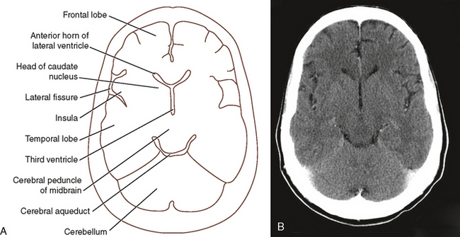
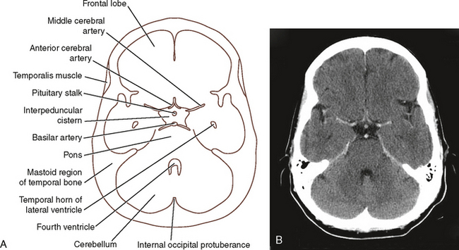
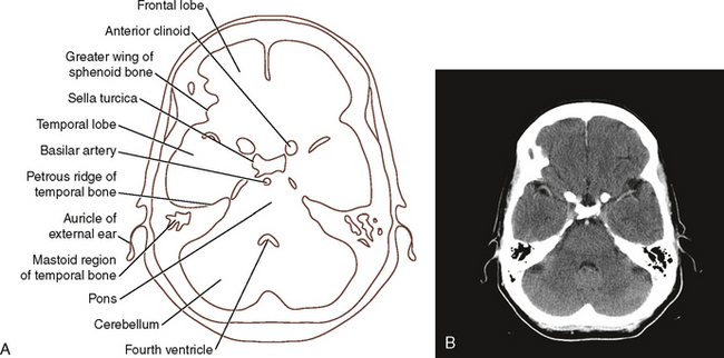
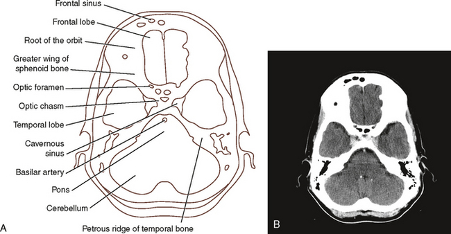
30 CT uses x-rays to generate images, so the various shades on the images correspond to the gray scale that radiographers are accustomed to seeing. Bones and other dense materials are white, whereas air and lower density materials are closer to black. Fat, muscle, and organs are represented with various shades of gray. Hounsfield units or CT numbers represent the scale of white to black that is used in CT imaging. Lower numbers represent anatomic structures that are more easily penetrated by the x-ray and appear closer to black on the image. Higher numbers are related to more radiopaque structures and are lighter gray or white on the image. Similar to routine radiographs, blood vessels and organs of the digestive system are not easily distinguishable from other structures. To be able to identify these structures more accurately, patients are frequently given a radiopaque contrast medium. Intravascular contrast medium highlights vessels, making them appear radiopaque and whiter on the image. To visualize the gastrointestinal system, patients may be given a contrast agent by mouth or via the rectum. For a full description of CT fundamentals, the reader is referred to Chapter 31. MRI uses magnetic fields and radiofrequencies to generate images. Anatomic structures are represented on the image with regard to the signal generated from their protons. Structures that produce a strong signal are generally lighter gray or white on the image, and structures that do not generate a strong signal tend to be darker on the image. The signal generated by these structures depends on many things, including the strength of the magnetic fields and the characteristics of the radiofrequencies used. Contrast medium may also be used when performing MRI to change the signal intensity of particular anatomic structures. Gadolinium, air, and fluid may be used as contrast agents depending on the organ of interest and the imaging sequences employed. MRI is discussed in depth in Chapter 32. Fig. 30-1 is a cadaveric image that can be used to distinguish bone, muscle, and other soft tissue structures. Referring to this image should be helpful in identifying the sometimes confusing shadows on the images. The head can be thought of systematically as being composed of the skull, central nervous system structures, various sensory organs, cranial blood supply, and associated cranial and facial muscles. The bones of the skull are categorized as the 8 cranial bones and the 14 facial bones. The cranial bones include the frontal, occipital, and two parietal bones that surround and protect the external surface of the brain. The other four cranial bones include the ethmoid, sphenoid, and two temporal bones. The frontal bone forms the anterior surface of the skull, with a vertical portion that corresponds to the forehead and a horizontal portion that forms the roof of the orbits. Between the inner and outer layers of the vertical portion of the frontal bone, just superior to the level of the eyes, are the paired frontal paranasal sinuses. The vertex (superiormost portion) of the skull is formed by the paired parietal bones. These roughly square-shaped bones articulate with the frontal bone at the coronal suture, with the temporal bones at the squamosal sutures, with the occipital bone at the lambdoidal suture, and with each other at the sagittal suture. In the center of the base of the skull is the sphenoid bone. This bone is sometimes referred to as the anchor bone of the cranium because it articulates with all of the cranial bones. Thinking of this bone as being composed of a body, two sets of wings, and a pterygoid portion is helpful. The body is the central portion of the bone and contains the easily identifiable landmark known as the sella turcica. The sella turcica forms a cup-shaped depression that surrounds and protects the pituitary gland. The anterior surface of the sella is called the tuberculum sellae, and the posterior portion is called the dorsum sellae. Two posterior clinoid processes project from the superior edge of the dorsum sellae and are attachments for dura mater partitions. Within the body of the sphenoid and inferior to the sella turcica are the paired sphenoidal paranasal sinuses. The lesser wings of the sphenoid are triangular ridges of bone found posterior to the orbital plates of the frontal bone. Directly inferior to the medial edge of each lesser wing is the optic canal, which transmits the optic nerve (cranial nerve II). The larger, greater wings support the temporal lobes of the cerebrum and extend from the body to the external surface of the skull. The pterygoid processes project inferiorly from the body of the sphenoid and form the posterior walls of the nasal cavity. The structures of the central nervous system within the skull include the cerebrum, brainstem, and cerebellum. The cerebrum is the largest of these structures and is divided by the longitudinal fissure into two hemispheres. The hemispheres are connected to each other via a white matter tract called the corpus callosum. This arch-shaped structure is divided into the anterior genu, central body, and posterior splenium. Each cerebral hemisphere is divided into lobes that are named for the most adjacent cranial bone: frontal, parietal, temporal, and occipital. An additional lobe called the insula is buried deep to the temporal lobe. The cerebrum is thrown into numerous folds called gyri, which are separated by small fissures called sulci. The outer surface of the cerebrum consists of a thin layer of gray matter. The central portion of this part of the brain is mainly white matter (formed by myelinated nerve fibers). These fibers, referred to as the corona radiata, connect the gray matter of the cortex to deeper gray matter nuclei deep within each hemisphere. The buried gray matter centers are called basal nuclei or basal ganglia and include the claustrum, putamen, globus pallidus, and caudate nucleus. White matter tracts, or capsules, are found between these gray matter structures. Other gray matter structures found within the central cerebrum include the thalamus and hypothalamus. These form the walls of the third ventricle. Many of these gray and white matter structures are seen on the cadaveric section in Fig. 30-1. Many muscles are associated with the face, only a few of which are referred to in the following sections. The temporalis muscle is found on the external surface of the squamous portion of the temporal bone. Its inferior attachment is to the coronoid process of the mandible. On the external surface of the mandibular rami are the masseter muscles, and on the internal surface of the rami are the pterygoid muscles. These muscles all are associated with moving the mandible and with swallowing. A lateral skull radiograph is used here for localization of the imaging plane in this section (Fig. 30-2, A), and a sagittal MR image (Fig. 30-2, B) is used for localization of MRI cross sections of the brain. CT imaging for the cranium may be performed with the gantry parallel to or angled 15 to 20 degrees to the orbitomeatal line. Angling the gantry of the CT scanner allows for imaging of the brain without excess radiation to the eyes. MRI of the cranium generally results in images that are parallel to the orbitomeatal or infraorbitomeatal plane. More details on patient positioning for CT are provided in Chapter 31, and information on patient positioning for MRI is provided in Chapter 32. Because the imaging planes may be different for CT and MRI, some variation exists in the anatomic structures visualized on corresponding illustrations in this section. Seven identifying lines represent the approximate levels for each of the labeled images for this region. The cranial CT image seen in Fig. 30-3 represents a CT slice obtained through the frontal and parietal bones, and Fig. 30-4 is a corresponding MR image. The cortex, or outer layer of gray matter, can be differentiated from the deeper white matter. The numerous gyri, or convolutions, and sulci are shown and are surrounded by the darker appearing CSF in the subarachnoid space. The cerebral hemispheres are separated by the longitudinal cerebral fissure. Invaginated in this fissure is a fold of dura mater, the falx cerebri. The superior sagittal sinus, which passes through the superior margin of the falx, follows the contour of the superior skull margin. In cross section, the anterior and posterior aspects of this sinus can normally be seen in the midline deep to the bony plates when the patient has been given an intravenous contrast agent and appear as triangular expansions near the bones easily seen on MR images. Two of the five cerebral lobes are seen (frontal and parietal). The corona radiata is the central tract of white matter in the cerebrum and is darker than the cortex on the CT image. This section passes through the superiormost portion of the corpus callosum, which separates the anterior and posterior portions of the falx cerebri. Fig. 30-3 A, Line drawing of CT section. B, CT image representing anatomic structures located at level A in Fig. 30-2, A. Fig. 30-5 is an axial CT slice through the superior portions of the lateral ventricles; Fig. 30-6 is the corresponding MR image. Visualized bony structures on the CT scan include the frontal bone and the two parietal bones. The falx cerebri is seen within the longitudinal fissure. The frontal lobes and parietal lobes of the cerebrum are shown. In the center of each image, the lateral ventricles are easily seen because of the dark appearance of the CSF circulating within each. In the posterior portions of the ventricles, the contrast-filled capillary network of the choroid plexuses also is visualized. A thin membrane called the septum pellucidum can be seen separating the ventricles. The corpus callosum is an arch-shaped structure; in cross section at this level, only the anterior genu and the posterior splenium can be seen. The caudate nuclei lie along the lateral surfaces of the ventricles and tend to follow their curves. Because these nuclei are composed of gray matter, they are the same shade of gray as the cortex on MR images (see Fig. 30-6). Several contrast-filled vascular structures are visible. The anterior cerebral arteries lie within the longitudinal fissure just anterior to the genu of the corpus callosum. A few branches of the middle cerebral arteries are seen near the lateral aspect of the skull on the CT scan. The anterior and posterior portions of the superior sagittal sinus are seen in the periphery of the falx cerebri. The inferior sagittal sinus lies in the internal edges of the falx. The thin strips of muscle seen on the external surface of the frontal bone correspond to the superior edges of the temporalis muscles. Fig. 30-5 A, Line drawing of CT section. B, CT image representing structures located at level B in Fig. 30-2, A. The axial sections through the mid-portion of the cerebrum show many of the central structures of the cerebral hemispheres (Fig. 30-7 is a CT image, and Fig. 30-8 is an MR image). Images at this level pass through the frontal bone, greater wing of the sphenoid, and squamous portion of the temporal bones. The posterior portion of the skull comprises the top portion (squamous portion) of the occipital bone at this level. The falx cerebri is shown within the longitudinal fissure, with the superior sagittal sinus best shown in the midline of the anterior and posterior margins of this membrane. In the CT image, the genu of the corpus callosum is found between the anterior horns of the lateral ventricles; however, the posterior portion of this slice is inferior to the level of the splenium. The MR image shows the genu and the splenium. At this level, the MR image shows the frontal, temporal, and occipital lobes along with the insula (fifth lobe or island of Reil), which is deep to the temporal lobe at the lateral fissure. Because of its orientation, the CT image shows the insula, frontal, and temporal lobes; the posterior aspect of the skull in this image is occupied by the cerebellum. Fig. 30-7 A, Line drawing of CT section. B, CT image representing anatomic structures located at level C in Fig. 30-2, A. The anterior and temporal horns of the lateral ventricles are seen on the CT scan, whereas the anterior and posterior horns are visible on the MR image. Within each posterior horn is a portion of the choroid plexus, which appears bright owing to the presence of contrast medium in the capillaries. The heads of the caudate nuclei lie along the external surfaces of the anterior horns of each lateral ventricle. Several areas of gray matter can be discerned on the CT image deep within the white matter of the cerebrum and constitute the basal nuclei. The MR image contrast has been enhanced so that the deep gray matter structures can be seen on it as well. The major components of the basal nuclei seen at this level are (from lateral to medial) the claustrum, lentiform nucleus (composed of the putamen and globus pallidus), and caudate nucleus. The lentiform nucleus is separated from the caudate nucleus and thalamus by a tract of white matter known as the internal capsule. These sections pass through the superior portion of the midline third ventricle. The thalamus, which serves as a central relay station for sensory impulses to the cerebral cortex, forms its lateral walls. The plane of the CT image passes through the structures of the midbrain. The anterior portions of the midbrain include the cerebral peduncles (white matter tracts that connect the cerebrum and the midbrain). The dark circular area at the posterior edge of the midbrain is the CSF-filled cerebral aqueduct. This passage connects the third and fourth ventricles and allows the circulation of CSF. A contrast-enhanced vessel, the great cerebral vein, is found just posterior to the third ventricle and the splenium of the corpus callosum on the MR image. It passes through the upper portion of the superior cistern. The pineal gland is also found in this cistern but is not clearly visualized in either image. This is an important radiographic landmark because of its tendency to calcify in adults. Branches of the middle cerebral artery are visible within the lateral fissures, and the anterior cerebral arteries can be seen in the anterior portion of the longitudinal fissure on the MR image. Fig. 30-9 is a CT image that passes through the frontal lobe, pons, and cerebellum; Fig. 30-10 is the MR image, which passes through the superior portions of the orbits, the midbrain, and the occipital lobes. Bony structures visible in the CT image include the frontal bone, the temporal bones, and the occipital bone. Within the temporal bones, the black air-filled structures represent the mastoid air cells. The internal protrusion of bone in the center of the occipital bone is the internal occipital protuberance. The area of signal void between the eyes on the MR image corresponds to the frontal sinuses. Frontal and temporal lobes of the cerebrum are shown on the CT image, whereas the frontal, temporal, and occipital lobes of the cerebrum and the insula are shown on the MR image. The CT scan passes just inferior to the midbrain, and the MR image passes through the level of the midbrain. The large dark area in the center of the CT image is the interpeduncular cistern. This is an enlarged area in the subarachnoid space containing CSF. The optic chiasm and the circle of Willis normally lie within the interpeduncular cistern. The pituitary stalk and some of the vessels that contribute to the circle of Willis are visible on this image. The pons lies posterior to the cistern. The cerebellum lies within the posterior fossa of the skull between the pons and the occipital bone. The large dark region between the pons and cerebellum is the CSF-filled fourth ventricle. The temporalis muscles are seen on the external surfaces on either side of the cranium. On the MR image, the cerebral peduncles form the anterior portions of the midbrain, and the corpora quadrigemina forms the posterior portion. The small black circle anterior to the colliculi is the CSF-filled cerebral aqueduct. Posterior to the midbrain is the cerebellum, which is surrounded by the tentorium cerebelli. The dark region anterior to the midbrain is the interpeduncular cistern, and the region posterior to the midbrain is the superior cistern. On the MR image, within the interpeduncular cistern, the optic tracts, hypothalamus, inferior portion of the third ventricle, and mammillary bodies can be seen. Several important vascular structures can be seen at this level. Fig. 30-9 A, Line drawing of CT section. B, CT image representing anatomic structures located at level D in Fig. 30-2, A. The CT image is just superior to the internal carotid arteries and shows the origins of the left anterior and middle cerebral arteries, at the anterior edge of the interpeduncular cistern. The anterior cerebral arteries pass from their origin toward the longitudinal fissure in the midline of the brain; the middle cerebral arteries course from their origins toward the lateral fissures. The CT image also shows the bifurcation of the basilar artery into the two posterior cerebral arteries. These vessels can be seen just anterior to the pons. The circle of Willis is an important vascular structure found in this region of the brain. Although it does not lie in the same plane as the imaging plane, much of the circle can be seen on the MR image. The bright anterior vascular structures represent the bifurcation of the internal carotid arteries into the anterior and middle cerebral arteries. The posterior cerebral arteries are seen here originating between the cerebral peduncles. The posterior and anterior communicating arteries are not seen in this image because they are not at the same level as the other vessels. The posterior portion of the superior sagittal sinus is located near the internal occipital protuberance; the straight sinus can be seen in the edge of the tentorium cerebelli, just posterior to the cerebellum on the MR image. Fig. 30-11 is a CT image through the sella turcica and the posterior fossa. The MR image (Fig. 30-12) passes through the center of the orbits, the tops of the ears, the pituitary and center of the sella turcica, and the cerebellum. The MR image shows the nasal bones, visible in the anterior skull. Between the eyes the ethmoidal sinuses are seen, and the cribriform plate of the ethmoid bone is seen. The sphenoidal sinuses lie posterior to the ethmoidal sinuses. The sella turcica and dorsum sellae are seen surrounding the pituitary gland. Several cranial bones are visible on the CT scan. The anterior clinoids of the sella turcica and the greater wings of the sphenoid are seen. The roof of the sella is formed by the lesser wings, anterior clinoids, and posterior clinoids. The temporal bone constitutes most of the lateral portions of the skull, and the petrous ridges can be seen on the CT image extending toward the median sagittal plane. The black air spaces near the lateral aspect of the petrous portions of these bones correspond to mastoid air cells, and the air spaces farther medial are associated with the internal structures of the ear. On the CT image, the frontal and temporal lobes of the cerebrum are visible, along with the pons and cerebellum. The dark region between the sella turcica and the pons is the pontine cistern, filled with CSF. The lower region of the fourth ventricle is seen between the pons and the cerebellum. On the MR image, both globes are visible within the orbits. Rectus muscles lie along the medial and lateral walls of each. The optic nerves are seen in the centers of the posterior orbits passing from the eyes toward the brain via the optic canal. The temporal lobes are found lateral to the sella turcica, resting in the middle cranial fossa. The pons lies posterior to the sella, and the cerebellum is seen filling the posterior cranial fossa. The edges of the tentorium cerebelli can be seen faintly between the temporal lobes and the cerebellum. The dark region anterior to the pons corresponds to the CSF-filled pontine cistern in which the contrast-filled basilar artery is easily visualized on both the CT and MR images. The dark region between the pons and the cerebellum is the superior region of the fourth ventricle. On the CT image, the contrast-filled basilar artery lies between the sella and the pons. At this level in the MR image, the internal carotid arteries lie lateral to the body of the sphenoid bone in an almost horizontal orientation. The confluence of sinuses can be seen just anterior to the internal occipital protuberance on the MR image. The confluence is the region where the superior sagittal sinus and the straight sinus meet the transverse sinuses. The transverse sinuses are seen on the MR image at this level. On the external surface of the skull in both images, the temporalis muscles lie along the temporal bones. The auricle, or cartilaginous portion of each ear, lies external to the temporal bone. Fig. 30-11 A, Line drawing of CT section. B, CT image representing anatomic structures located at level E in Fig. 30-2, A. The sectional images through the lower cranium show the inferior portions of the cerebrum, brainstem, cerebellum, and associated major skeletal structures (Fig. 30-13 is a CT image, and Fig. 30-14 is an MR image). The CT image shows the frontal sinuses and the roofs of the orbits. The greater and lesser wings of the sphenoid bone are shown. The optic foramina (canals) can be seen between the greater and lesser wings. The optic chiasm and cavernous sinus can be seen posterior to the optic foramen. The petrous and mastoid portions of the temporal bones are shown dividing the middle and posterior cranial fossae. The maxilla, maxillary sinuses, and nasal bones are seen in the anterior skull on the MR image (note the mass within the right maxillary sinus). The zygomatic bones form the lateral walls of the orbits, and the maxillae form the medial walls. The perpendicular plate and vomer form the bony nasal septum seen in the center of the nasal cavity. Posterior to the nasal cavity, the sphenoidal sinuses are seen between the lower aspects of the greater wings. Both petrous ridges extend toward the midline; these are seen as dark areas on the MR image because of the lack of signal from this dense region of bone. Extending into the right petrous ridge is the external auditory canal. Just anterior to the canal is the condyle of the mandible resting in the mandibular fossa. Mastoid air cells lie posterior to the external acoustic meatus. Fig. 30-13 A, Line drawing of CT section. B, CT image representing anatomic structures located at level F in Fig. 30-2, A.
SECTIONAL ANATOMY FOR RADIOGRAPHERS
Overview
Cranial Region
![]()
Stay updated, free articles. Join our Telegram channel

Full access? Get Clinical Tree


Radiology Key
Fastest Radiology Insight Engine