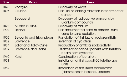
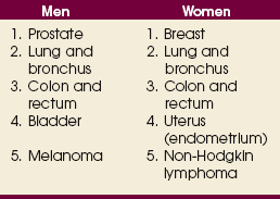
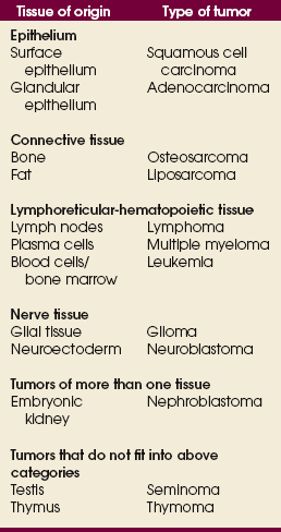
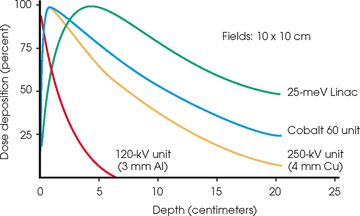
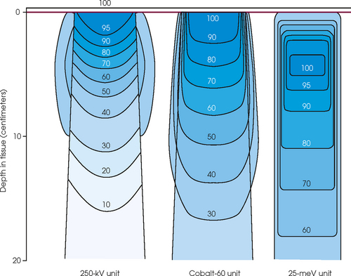
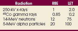
RADIATION ONCOLOGY

36


RADIATION ONCOLOGY


Principles of Radiation Oncology


image


Radiation oncology,* or radiation therapy, is one of three principal modalities used in the treatment of cancer. The others are surgery and chemotherapy. In radiation therapy for malignancies, tumors or lesions are treated with cancericidal doses of ionizing radiation as prescribed by a radiation oncologist, a physician who specializes in the treatment of malignant disease with radiation. The goals of the treatment are to deliver a cancericidal dose of radiation precisely to the tumor, limiting as much as possible the dose of radiation received by normal, noncancerous tissues. These dual tasks make this form of treatment complex and often challenging. Input from all members of the radiation oncology team is crucial in developing the optimal treatment plan or approach for a patient.


Cancer treatment requires a multidisciplinary approach. First, diagnostic radiologic studies such as radiographs, computed tomography (CT) scans, magnetic resonance imaging (MRI), positron emission tomography (PET) scans, and sonograms are obtained to acquire information about the location and anatomic extent of the tumor. Second, a tissue specimen (biopsy) is removed surgically. A pathologist examines the tissue to determine whether the lesion is cancerous. When cancer is diagnosed, the plan for the best treatment is determined through consultation with various oncology specialists (e.g., surgical oncologist, radiation oncologist, medical oncologist).


Although radiation oncology may be used as the only method of treatment for malignant disease, a more common approach is to use radiation in conjunction with surgery or chemotherapy or both. Some patients with cancer may be treated only with surgery or chemotherapy; however, approximately 75% of all diagnosed cancer patients are treated with radiation. The choice of treatment can depend on many patient variables, such as the patient’s overall physical and emotional condition, the histologic type of the disease, and the extent and anatomic position of the tumor. If a tumor is small and its margins are well defined, a surgical approach alone may be prescribed. If the disease is systemic, a chemotherapeutic approach may be chosen. Most tumors exhibit degrees of size, invasion, and spread, however, and require variations in the treatment approach that are likely to include radiation treatments administered as an adjunct to or in conjunction with surgery or chemotherapy.


Radiation is generally used after surgery when a patient is deemed to be at high risk for tumor recurrence in the surgical bed. The risk of recurrence is considered to be increased in the following situations:


Radiation can be used as definitive (primary) cancer treatment or adjuvant treatment (i.e., in combination with another form of therapy). It can also be used for palliation.


Radiation treatments most often are delivered on a daily basis, Monday through Friday, for 2 to 8 weeks. The length of time and the total dose of radiation delivered depend on the type of cancer being treated and the purpose of treatment (cure or palliation). Prescribed dosages of radiation can range from 2000 centigray (cGy) for palliation to 8000 cGy for curative intent (total doses). The delivery of a small amount of radiation per day (180 to 200 cGy) for a certain number of treatments, instead of one large dose, is termed fractionation. Because these smaller doses of radiation are more easily tolerated by normal tissue, fractionation can help minimize the acute toxic effect a patient experiences during treatment and the possible long-term side effects of treatment.


The precision and accuracy necessary to administer high doses of radiation to tumors while not harming normal tissue require the combined effort of all members of the radiation oncology team. Members of this team include the radiation oncologist, a physicist, dosimetrists, radiation therapists, and oncology nurses.


The radiation oncologist prescribes the quantity of radiation and determines the anatomic region or regions to be treated. The medical physicist is responsible for calibration and maintenance of the radiation-producing equipment. The physicist also advises the physician about dosage calculations and complex treatment techniques. The medical dosimetrist devises a plan for delivering the treatments in a manner to meet best the physician’s goals of irradiating the tumor while protecting vital normal structures. The radiation therapist is responsible for obtaining radiographs or CT scans that localize the area to be treated, administering the treatments, keeping accurate records of the dose delivered each day, and monitoring the patient’s physical and emotional well-being. Educating patients about potential radiation side effects and assisting patients with the management of these side effects are often the responsibilities of the oncology nurse.


The duties and responsibilities of the radiation therapist are more thoroughly described elsewhere in this chapter. In addition, more information is provided about the circumstances in which radiation is used to treat cancer. The steps necessary to prepare a patient for treatment are also described. These steps include (1) simulation, (2) development of the optimal treatment plan in dosimetry, and (3) treatment delivery. Current techniques and future trends are also discussed.


Historical Development


Ionizing radiation was originally used to obtain a radiographic image of internal anatomy for diagnostic purposes. The resultant image depended on many variables, including the energy of the beam, the processing techniques, the material on which the image was recorded, and, most importantly, the amount of energy absorbed by the various organs of the body. The transfer of energy from the beam of radiation to the biologic system and the observation of the effects of this interaction became the foundation of radiation oncology.


Two of the most obvious and sometimes immediate biologic effects observed during the early diagnostic procedures were epilation (loss of hair) and erythema (reddening of the skin). Epilation and erythema resulted primarily from the great amount of energy absorbed by the skin during radiographic procedures. These short-term, radiation-induced effects afforded radiographic practitioners an opportunity to expand the use of radiation to treat conditions ranging from relatively benign maladies such as hypertrichosis (excessive hair), acne, and boils to grotesque and malignant diseases such as lupus vulgaris and skin cancer.


Ionizing radiation was first applied for the treatment of a more in-depth lesion on January 29, 1896, when Grubbé is reported to have irradiated a woman with carcinoma of the left breast. This event occurred only 3 months after the discovery of x-rays by Röntgen (Table 36-1). Although Grubbé neither expected nor observed any dramatic results from the irradiation, the event is significant simply because it occurred.


The first reported curative treatment using ionizing radiation was performed by Skinner in New Haven, Connecticut, in January 1902. Skinner treated a woman who had a diagnosed malignant fibrosarcoma. Over the next 2 years and 3 months, the woman received 136 applications of the x-rays. In April 1909, 7 years after initial application of the radiation, the woman was free of disease and considered “cured.”


As data were collected, the interest in radiation therapy increased. More sophisticated equipment, a greater understanding of the effects of ionizing radiation, an appreciation for time-dose relationships, and numerous other related medi-cal breakthroughs gave impetus to the interest in radiation therapy that led to the evolution of a distinct medical specialty—radiation oncology.


Cancer


Cancer is a disease process that involves an unregulated, uncontrolled replication of cells; put more simply, the cells do not know when to stop dividing. These abnormal cells grow without regard to normal tissue. They invade adjacent tissues, destroy normal tissue, and create a mass of tumor cells. Cancerous cells can spread further by invading the lymph or blood vessels that drain the area. When tumor cells invade the lymphatic or vascular system, they are transported by that system until they become caught or lodged within a lymph node or an organ such as the liver or lungs, where secondary tumors form. The spread of cancer from the original site to different, remote parts of the body is termed metastasis. When cancer has spread to a distant site via blood-borne metastasis, the patient is considered incurable. Early detection and diagnosis are the keys to curing cancer.


Cancer was diagnosed in an estimated 1,479,350 individuals in the United States in 2009. This number does not include basal and squamous cell skin cancers, which have high cure rates. These types of cancer are the most common malignant diseases, with more than 1.3 million cases diagnosed in 2000. One in two men will develop or die of cancer in their lifetime. Slightly more than one in three women will develop or die of cancer in their lifetime. Although cancer can occur in persons of any age, it is diagnosed in most patients after age 55 years.


The most common cancers that occur in the United States are lung, prostate, breast, and colorectal cancer. Prostate cancer is the most common malignancy in men, and breast cancer is the most common malignancy in women. The second and third most common cancers in men and women are lung and colorectal cancer (Table 36-2).


Cancer is second only to heart disease as the leading cause of death in the United States. Lung cancer is the leading cause of cancer deaths for men and women. In 2009 an estimated 30% of cancer deaths in men and 26% in women were due to lung cancer. The next most common causes of cancer death are prostate cancer and breast cancer, which account for 9% and 15% of cancer deaths in the United States.


RISK FACTORS


External factors


Many factors can contribute to a person’s potential for the development of a malignancy. These factors can be external exposure to chemicals, viruses, or radiation within the environment or internal factors such as hormones, genetic mutations, and disorders of the immune system. Cancer commonly is the result of exposure to a carcinogen, which is a substance or material that causes cells to undergo malignant transformation and become cancerous. Some known carcinogenic agents are listed in Table 36-3. Cigarettes and other tobacco products are the principal cause of cancers of the lung, esophagus, oral cavity and pharynx, and bladder. Cigarette smokers are 10 times more likely to develop lung cancer than nonsmokers. Occupational exposure to chemicals such as chromium, nickel, or arsenic can also cause lung cancer. A person who smokes and works with chemical carcinogens is at even greater risk for developing lung cancer than a nonsmoker. In other words, risk factors can have an additive effect, acting together to initiate or promote the development of cancer.


Another carcinogen is ionizing radiation. It was responsible for the development of osteogenic sarcoma in radium-dial painters in the 1920s and 1930s, and it caused the development of skin cancers in pioneer radiologists. Early radiation therapy equipment used in the treatment of cancer often induced a second malignancy in the bone. The low-energy x-rays produced by this equipment were within the photoelectric range of interactions with matter, resulting in a 3:1 preferential absorption in bone compared with soft tissue. Some patients with breast cancer who were irradiated developed an osteosarcoma of their ribs after a 15- to 20-year latency period. With advances in diagnostic and therapeutic equipment and improved knowledge of radiation physics, radiobiology, and radiation safety practices, radiation-induced malignancies have become relatively uncommon, although the potential for their development still exists. In keeping with standard radiation safety guidelines, any dose of radiation, no matter how small, significantly increases the chance of a genetic mutation.


Internal factors


Internal factors are causative factors over which persons have no control. Genetic mutations on individual genes and chromosomes have been identified as predisposing factors for the development of cancer. Mutations can be sporadic or hereditary, as in colon cancer. Chromosomal defects have also been identified in other cancers, such as leukemia, Wilms tumor, retinoblastoma, and breast cancer. Because of their familial pattern of occurrence, breast, ovarian, and colorectal cancer are three major areas currently under study to obtain earlier diagnosis, which increases the cure rate. Patients with a family history of breast or ovarian cancer can be tested to see whether they have inherited the altered BRCA-1 and BRCA-2 genes. Patients with these altered genes are at a significantly higher risk for developing breast and ovarian cancer. Women identified as carriers of the altered genes can benefit from more intensive and early screening programs in which breast cancer may be diagnosed at a much earlier and more curable stage. These patients also have the option of prophylactic surgery to remove the breasts or ovaries. Some women still develop cancer, however, in the remaining tissue after surgery.


TISSUE ORIGINS OF CANCER


Cancers may arise in any human tissue. Tumors are usually categorized under six general headings according to their tissue of origin (Table 36-4). Of cancers, 90% arise from epithelial tissue and are classified as carcinomas. Epithelial tissue lines the free internal and external surfaces of the body. Carcinomas are subdivided further into squamous cell carcinomas and adenocarcinomas based on the type of epithelium from which they arise. A squamous cell carcinoma arises from the surface (squamous) epithelium of a structure. Examples of surface epithelium include the oral cavity, pharynx, bronchus, skin, and cervix. An adenocarcinoma is a cancer that develops in glandular epithelium such as in the prostate, colon and rectum, lung, breast, or endometrium.


To facilitate the exchange of patient information from one physician to another, a system of classifying tumors based on anatomic and histologic considerations was designed by the International Union Against Cancer and the American Joint Committee for Cancer (AJCC) Staging and End Results Reporting. The AJCC TNM classification (Table 36-5) describes a tumor according to the size of the primary lesion (T), the involvement of the regional lymph nodes (N), and the occurrence of metastasis (M).


Theory


The biologic effectiveness of ionizing radiation in living tissue depends partially on the amount of energy that is deposited within the tissue and partially on the condition of the biologic system. The terms used to describe this relationship are linear energy transfer (LET) and relative biologic effectiveness (RBE).


LET values are expressed in thousands of electron volts deposited per micron of tissue (keV/μm) and vary depending on the type of radiation being considered. Because of their mass and possible charge, particles tend to interact more readily with the material through which they are passing and have a greater LET value. A 5-MeV alpha particle has an LET value of 100 keV/mm in tissue; nonparticulate radiations such as 250-kilovolt (peak) (kVp) x-rays and 1.2-MeV gamma rays have much lower LET values: 2.0 keV/mm and 0.2 keV/mm.


RBE values are determined by calculating the ratio of the dose from a standard beam of radiation to the dose required of the radiation beam in question to produce a similar biologic effect. The standard beam of radiation is 250-kVp x-rays, and the ratio is set up as follows:


image


As the LET increases, so does the RBE. RBE and LET values are listed in Table 36-6.


The effectiveness of ionizing radiation on a biologic system depends not only on the amount of radiation deposited but also on the state of the biologic system. One of the first laws of radiation biology, postulated by Bergonié and Tribondeau, stated in essence that the radiosensitivity of a tissue depends on the number of undifferentiated cells in the tissue, the degree of mitotic activity of the tissue, and the length of time that cells of the tissue remain in active proliferation. Although exceptions exist, the preceding is true in most tissues. The primary target of ionizing radiation is the DNA molecule, and the human cell is most radiosensitive during mitosis. Current research tends to indicate that all cells are equally radiosensitive; however, the manifestation of the radiation injury occurs at different time frames (i.e., acute vs. late effects).


Because tissue cells are composed primarily of water, most of the ionization occurs with water molecules. These events are called indirect effects and result in the formation of free radicals such as OH, H, and HO2. These highly reactive free radicals may recombine with no resultant biologic effect, or they may combine with other atoms and molecules to produce biochemical changes that may be deleterious to the cell. The possibility also exists that the radiation may interact with an organic molecule or atom, which may result in the inactivation of the cell; this reaction is called the direct effect. Because ionizing radiation is nonspecific (i.e., it interacts with normal cells as readily as with tumor cells), cellular damage occurs in normal and abnormal tissue. The deleterious effects are greater in the tumor cells, however, because a greater percentage of these cells are undergoing mitosis; tumor cells also tend to be more poorly differentiated. In addition, normal cells have a greater capability for repairing sublethal damage than tumor cells. Greater cell damage occurs to tumor cells than to normal cells for any given increment of dose. The effects of the interactions in either normal or tumor cells may be expressed by the following descriptions:


The greater the number of interactions that occur, the greater the possibility of cell death.


The preceding information leads to a categorization of tumors according to their radiosensitivity:


Many concepts that originate in the laboratory have little practical application, but some are beginning to influence the selection of treatment modalities and the techniques of radiation oncology. As cellular function and the effects of radiation on the cell are increasingly understood, attention is being focused on the use of drugs, or simply oxygen, to enhance the effectiveness of radiation treatments.


Technical Aspects


EXTERNAL-BEAM THERAPY AND BRACHYTHERAPY


Two major categories for the application of radiation for cancer treatment are external-beam therapy and brachytherapy. For external-beam treatment, the patient lies underneath a machine that emits radiation or generates a beam of x-rays. This technique is also called teletherapy, or long-distance treatment. Most cancer patients are treated in this fashion. Some patients may also be treated with brachytherapy, a technique in which the radioactive material is placed within the patient.


The theory behind brachytherapy is to deliver low-intensity radiation over an extended period to a relatively small volume of tissue. The low-intensity isotopes are placed directly into a tissue or cavity depositing radiation only a short distance, covering the tumor area but sparing surrounding normal tissue. This technique allows a higher total dose of radiation to be delivered to the tumor than is achievable with external-beam radiation alone. Brachytherapy may be accomplished in any of the following ways:


Most brachytherapy applications tend to be temporary in that the sources are left in the patient until a designated tumor dose has been attained. Two different brachytherapy systems exist. They are low-dose-rate (LDR) and high-dose-rate (HDR). LDR brachytherapy has been the standard system for many years. A low-activity isotope is used to deliver a dose of radiation at a slow rate of 40 to 500 cGy per hour. This therapy requires that a patient be hospitalized for 3 to 4 days until the desired dose is delivered.


HDR systems are becoming the more standard method of brachytherapy. This system uses a high-activity isotope capable of delivering greater than 1200 cGy per hour. The HDR system allows the prescribed dose to be delivered over minutes, which means this treatment can occur on an outpatient basis. Gynecologic tumors are one of the most common sites to be treated with LDR or HDR brachytherapy. Classic LDR systems use the isotopes cesium-137 for intracavity applications and iridium-192 for interstitial applications. HDR systems use a high-activity iridium-192 source.


Permanent implant therapy may also be accomplished. An example of a permanent implant nuclide is iodine-125 seeds. Permanent implant nuclides have half-lives of hours or days and are left in the patient essentially forever. The amount and distribution of the radionuclide implanted in this manner depends on the total dose that the radiation oncologist is trying to deliver. Early-stage prostate cancer is commonly treated with this technique. In most cases of brachytherapy implantation, the implant is applied as part of the patient’s overall treatment plan and may be preceded by or followed by additional external-beam radiation therapy or possibly surgery.


EQUIPMENT


Most radiation oncology departments use linear accelerators (linacs) as their main treatment unit. Following are treatment units that may be found in a radiation oncology department:


The dose depositions of these units are compared in Fig. 36-1.


The penetrability, or energy, of an x-ray or gamma ray totally depends on its wavelength: The shorter the wavelength, the more penetrating the photon; conversely, the longer the wavelength, the less penetrating the photon. A low-energy beam (≤120 kVp) of radiation tends to deposit all or most of its energy on or near the surface of the patient and is suitable for treating lesions on or near the skin surface. In addition, with the low-energy beam, a greater amount of absorption or dose deposition occurs in bone than in soft tissue.


A high-energy beam of radiation (≥1 MeV) tends to deposit its energy throughout the entire volume of tissue irradiated, with a greater amount of dose deposition occurring at or near the entry port than at the exit port. In this energy range, the dose is deposited about equally in soft tissue and bone. The high-energy (megavoltage) beam is most suitable for tumors deep beneath the body surface.


The skin-sparing effect, a phenomenon that occurs as the energy of a beam of radiation is increased, is valuable from a therapeutic standpoint. In the superficial and orthovoltage energy range, the maximum dose occurs on the surface of the patient, and deposition of the dose decreases as the beam traverses the patient. As the energy of the beam increases into the megavoltage range, the maximum dose absorbed by the patient occurs at some point below the skin surface. The skin-sparing effect is important clinically because the skin is a radiosensitive organ. Excessive dose deposition to the skin can damage the skin, requiring treatments to be stopped and compromising treatment to the underlying tumor. The greater the energy of the beam, the more deeply the maximum dose is deposited (Fig. 36-2).


Cobalt-60 units


The 60Co unit was the first skin-sparing machine. It replaced the orthovoltage unit in the early 1950s because of its greater ability to treat tumors located deeper within tissues. 60Co is an artificially produced isotope formed in a nuclear reactor by the bombardment of stable cobalt-59 with neutrons. 60Co emits two gamma-ray beams with an energy of 1.17 MeV and 1.33 MeV. The unit was known as a “workhorse” because it was extremely reliable, was mechanically simple, and had little downtime. It was the first radiation therapy unit to rotate 360 degrees around a patient. A machine that rotates around a fixed point, or axis, and maintains the same distance from the source of radiation is called an isocentric machine. All modern therapeutic units are isocentric machines. This type of machine allows the patient to remain in one position, lessening the chance for patient movement during treatment. Isocentric capabilities also assist in directing the beam precisely at the tumor while sparing normal structures.


Because 60Co is a radioisotope, it constantly emits radiation as it decays in an effort to return to a stable state. It has a half-life of 5.26 years (i.e., its activity is reduced by 50% at the 5.26 years). Because the source decays at a rate of 1% per month, the radiation treatment time must be adjusted, resulting in longer treatment times as the source decays.


The use of 60Co units has declined significantly since the 1980s, and 60Co is rarely used for conventional external-beam radiation therapy today. This decline has been basically attributed to the introduction of the more sophisticated linac, which has greater skin-sparing capabilities and more sharply defined radiation fields. The radiation beam, or field, from a 60Co unit also has large penumbra, which results in fuzzy field edges, another undesirable feature. 60Co is still used in radiation oncology as part of a special procedure called stereotactic radiosurgery. The treatment unit is called the Gamma Knife. The Gamma Knife consists of 192 to 201 60Co sources arranged in a hemispherical array with all sources converging at a single point (Fig. 36-3). The point where the beams converge forms a treatment area of 4 to 18 mm in diameter.


The Gamma Knife is primarily used to treat small benign or malignant lesions located deep within the brain employing an external rigidly fixed stereotactic head frame. The Gamma Knife does not involve surgery. It is called radiosurgery because the radiation is delivered in such a precise, focused manner that the lesion is ablated as if removed surgically. Adjacent normal tissues receive minimal radiation and are unharmed. The stereotactic head frame provides a coordinate system that allows the lesion to be three-dimensionally localized on MRI, CT scan, or angiography so that the radiation can be planned and targeted directly to the involved area. The Gamma Knife delivers a large dose of radiation in a single treatment to one or more areas in the brain. The types of conditions treated with the Gamma Knife include benign conditions such as acoustic neuromas, pituitary adenomas, arteriovenous malformations, and trigeminal neuralgia. Malignant lesions treated with the Gamma Knife include gliomas, meningiomas, chordoma, and solitary brain metastasis.


There are many advantages of Gamma Knife radiosurgery over conventional neurosurgery. First, the patient does not have to undergo an invasive surgical procedure. The procedure can be done as an outpatient or may require an overnight stay in the hospital. There is no major recuperation period after a Gamma Knife procedure. The cost of Gamma Knife radiosurgery is much less than the cost of neurosurgery. The Gamma Knife is considered a very effective treatment for small intracranial lesions. One disadvantage of the Gamma Knife is that it can be used only for intracranial lesions. Another disadvantage is that the effects of radiation on the lesion are not immediate but occur over a period of weeks.


Linear accelerators


Linacs are the most commonly used machines for cancer treatment. The first linac was developed in 1952 and first used clinically in the United States in 1956. A linac is capable of producing high-energy beams of photons (x-rays) or electrons in the range of 4 million to 35 million volts. These megavoltage photon beams allow a better distribution of dose to deep-seated tumors with better sparing of normal tissues than their earlier counterparts—the orthovoltage or 60Co units.

Stay updated, free articles. Join our Telegram channel

Mar 4, 2016 | Posted by in GENERAL RADIOLOGY | Comments Off on RADIATION ONCOLOGY

Full access? Get Clinical Tree

Get Clinical Tree app for offline access