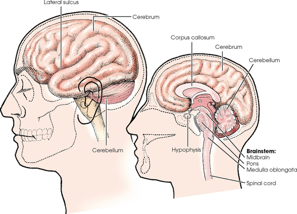
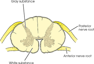
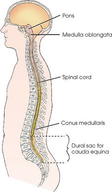
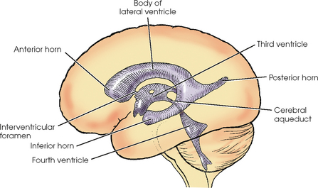
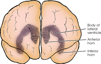
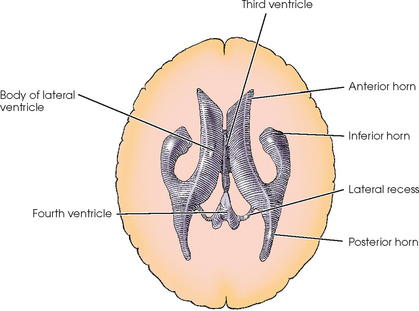
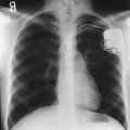
CENTRAL NERVOUS SYSTEM

24


CENTRAL NERVOUS SYSTEM


ANATOMY


image


For descriptive purposes, the central nervous system (CNS) is divided into two parts: (1) the brain,* which occupies the cranial cavity, and (2) the spinal cord, which is suspended within the vertebral canal.


Brain


The brain is composed of an outer portion of gray matter called the cortex and an inner portion of white matter. The brain consists of the cerebrum; cerebellum; and brainstem, which is continuous with the spinal cord (Fig. 24-1). The brainstem consists of the midbrain, pons, and medulla oblongata.


The cerebrum is the largest part of the brain and is referred to as the forebrain. Its surface is convoluted by sulci and grooves that divide it into lobes and lobules. The stemlike portion that connects the cerebrum to the pons and cerebellum is termed the midbrain. The cerebellum, pons, and medulla oblongata make up the hindbrain.


A deep cleft, called the longitudinal sulcus (interhemispheric fissure), separates the cerebrum into right and left hemispheres, which are closely connected by bands of nerve fibers, or commissures. The largest commissure between the cerebral hemispheres is the corpus callosum. The corpus callosum is a midline structure inferior to the longitudinal sulcus. Each cerebral hemisphere contains a fluid-filled cavity called a lateral ventricle. At the diencephalon, or second portion of the brain, the thalami surround the third ventricle. Inferior to the diencephalon is the pituitary gland, the master endocrine gland of the body. The pituitary gland resides in the hypophyseal fossa of the sella turcica.


The cerebellum, the largest part of the hindbrain, is separated from the cerebrum by a deep transverse cleft. The hemispheres of the cerebellum are connected by a median constricted area called the vermis. The surface of the cerebellum contains numerous transverse sulci that account for its cauliflowerlike appearance. The tissues between the curved sulci are called folia. The pons, which forms the upper part of the hindbrain, is the commissure or bridge between the cerebrum, cerebellum, and medulla oblongata. The medulla oblongata, which extends between the pons and spinal cord, forms the lower portion of the hindbrain. All the fiber tracts between the brain and spinal cord pass through the medulla.


Spinal Cord


The spinal cord is a slender, elongated structure consisting of an inner, gray, cellular substance, which has an H shape on transverse section, and an outer, white, fibrous substance (Figs. 24-2 and 24-3). The cord extends from the brain, where it is connected to the medulla oblongata at the level of the foramen magnum, to the approximate level of the space between the first and second lumbar vertebrae. The spinal cord ends in a pointed extremity called the conus medullaris (see Fig. 24-3). The filum terminale is a delicate fibrous strand that extends from the terminal tip and attaches the cord to the upper coccygeal segment.


In an adult, the spinal cord is 18 to 20 inches (46 to 50 cm) long and is connected to 31 pairs of spinal nerves. Each pair of spinal nerves arises from two roots at the sides of the spinal cord. The nerves are transmitted through the intervertebral and sacral foramina. Spinal nerves below the termination of the spinal cord extend inferiorly through the vertebral canal. These nerves resemble a horse’s tail and are referred to as the cauda equina. The spinal cord and nerves work together to transmit and receive sensory, motor, and reflex messages to and from the brain.


Meninges


The brain and spinal cord are enclosed in three continuous protective membranes called meninges. The inner sheath, called the pia mater (Latin, meaning “tender mother”), is highly vascular and closely adherent to the underlying brain and cord structure.


The delicate central sheath is called the arachnoid. This membrane is separated from the pia mater by a comparatively wide space called the subarachnoid space, which is widened in certain areas. These areas of increased width are called subarachnoid cisterns. The widest area is the cisterna magna (cisterna cerebellomedullaris). This triangular cavity is situated in the lower posterior fossa between the base of the cerebellum and the dorsal surface of the medulla oblongata. The subarachnoid space is continuous with the ventricular system of the brain and communicates with it through the foramina of the fourth ventricle. The ventricles of the brain and the subarachnoid space contain cerebrospinal fluid (CSF). CSF is the tissue fluid of the brain and spinal cord; it surrounds and cushions the structures of the CNS.


The outermost sheath, called the dura mater (Latin, meaning “hard mother”), forms the strong fibrous covering of the brain and spinal cord. The dura is separated from the arachnoid by the subdural space and from the vertebral periosteum by the epidural space. These spaces do not communicate with the ventricular system. The dura mater is composed of two layers throughout its cranial portion. The outer layer lines the cranial bones, serving as periosteum to their inner surface. The inner layer protects the brain and supports the blood vessels. The inner layer also has four partitions that provide support and protection for the various parts of the brain. One of these partitions, the falx cerebri, runs through the interhemispheric fissure and provides support for the cerebral hemispheres. The tentorium is a tent-shaped fold of dura that separates the cerebrum and cerebellum. Changes in the normal positions of these structures often indicate pathology. The dura mater extends below the spinal cord (to the level of the second sacral segment) to enclose the spinal nerves, which are prolonged inferiorly from the cord to their respective exits. The lower portion of the dura mater is called the dural sac. The cauda equina is enclosed by the dural sac.


Ventricular System


The ventricular system of the brain consists of four irregular, fluid-containing cavities that communicate with one another through connecting channels (Figs. 24-4 to 24-6). The two upper cavities are an identical pair and are called the right and left lateral ventricles. They are situated, one on each side of the midsagittal plane, in the inferior medial part of the corresponding hemisphere of the cerebrum.


Each lateral ventricle consists of a central portion called the body of the cavity. The body is prolonged anteriorly, posteriorly, and inferiorly into hornlike portions that give the ventricle an approximate U shape. The prolonged portions are known as the anterior, posterior, and inferior horns. Each lateral ventricle is connected to the third ventricle by a channel called the interventricular foramen or foramen of Monro, through which it communicates directly with the third ventricle and indirectly with the opposite lateral ventricle.


The third ventricle is a slitlike cavity with a quadrilateral shape. It is situated in the midsagittal plane just inferior to the level of the bodies of the lateral ventricles. This cavity extends anteroinferiorly from the pineal gland, which produces a recess in its posterior wall, to the optic chiasm, which produces a recess in its anteroinferior wall.


The interventricular foramina, one from each lateral ventricle, open into the anterosuperior portion of the third ventricle. The cavity is continuous posteroinferiorly with the fourth ventricle by a passage known as the cerebral aqueduct, or aqueduct of Sylvius.


The fourth ventricle is diamond-shaped and is located in the area of the hindbrain. The fourth ventricle is anterior to the cerebellum and posterior to the pons and the upper portion of the medulla oblongata. The distal, pointed the fourth ventricle is continuous with the central canal of the medulla oblongata. CSF exits the fourth ventricle into the subarachnoid space via the median aperture (foramen of Magendie) and the lateral apertures (foramen of Luschka).


Plain Radiographic Examination


Neuroradiologic assessment should begin with noninvasive imaging procedures. Radiographs of the cerebral and visceral cranium and the vertebral column may be obtained to show bony anatomy. In traumatized patients (see Chapter 8), radiographs are obtained to detect bone injury, subluxation, or dislocation of the vertebral column and to determine the extent and stability of the bone injury.


For a traumatized patient with possible CNS involvement, a cross-table lateral cervical spine radiograph should be obtained first to rule out fracture or misalignment of the cervical spine. Approximately two thirds of significant pathologic conditions affecting the spine can be detected on this initial image. Care must be taken to show the entire cervical spine adequately including the C7-T1 articulation. Employing the Twining (swimmer’s) method (see Chapter 8) may be necessary to show this anatomic region radiographically.


After the cross-table lateral radiograph has been checked and cleared by a physician, the following cervical spine projections should be obtained: anteroposterior (AP), bilateral AP oblique (trauma technique may be necessary), and AP to show the dens. A vertebral arch, or pillar image, of the cervical spine may provide additional information about the posterior portions of the cervical vertebrae (see Chapter 8). An upright lateral cervical spine radiograph may also be requested to show alignment of the vertebrae better and to assess the normal lordotic curvature of the spine.


Tomography may be used to supplement images of the spine for initial screening purposes. Tomography has been largely replaced by computed tomography (CT) in many institutions (see Chapter 31). Tomography may be employed to show long continuous areas of the spine. Disadvantages of tomography include the lack of soft tissue detail and the difficulty in positioning a traumatized patient for lateral tomographic radiographs.


Radiographs of the spine should always be obtained before myelography. Routine images of the vertebral column are helpful in assessing narrowed disk spaces because of degeneration of the disk, osteoarthritis, postoperative changes in the spine, and other pathologies of the vertebral column. Because the contrast agents used in myelography may obscure some anomalies, noncontrast spinal images complement the myelographic examination and often provide additional information.


Routine skull images should be obtained when the possibility of a skull fracture exists. In trauma patients, a cross-table lateral or upright lateral skull radiograph must be obtained to show air-fluid levels in the sphenoid sinus. In many instances, these air-fluid levels may be the initial indication of a basilar skull fracture. In addition, skull images are helpful in diagnosing reactive bone formation and general alterations in the skull resulting from various pathologic conditions, including Paget disease, fibrous dysplasia, hemangiomas, and changes in the sella turcica.


Myelography


Myelography (Greek, myelos,“marrow; the spinal cord”) is the general term applied to radiologic examination of the CNS structures situated within the vertebral canal. This examination is performed by introducing a nonionic, water-soluble contrast medium into the subarachnoid space by spinal puncture, most commonly at the L2-3 or L3-4 interspace or at the cisterna magna between C1 and the occipital bone. Injections into the subarachnoid space are termed intrathecal injections.


Most myelograms are performed on an outpatient basis, with patients recovering for approximately 4 to 8 hours after the procedure before being released to return home. In many parts of the United States, magnetic resonance imaging (MRI) (see Chapter 32) has largely replaced myelography. Myelography continues to be the preferred examination method for assessing disk disease in patients with contraindications to MRI, such as pacemakers or metallic posterior spinal fusion rods.


Myelography is employed to show extrinsic spinal cord compression caused by a herniated disk, bone fragments, or tumors and spinal cord swelling resulting from traumatic injury. These encroachments appear radiographically as a deformity in the subarachnoid space or an obstruction of the passage of the column of contrast medium within the subarachnoid space. Myelography is also useful in identifying narrowing of the subarachnoid space by evaluating the dynamic flow patterns of the CSF.


CONTRAST MEDIA


A non–water-soluble, iodinated ester (iophendylate [Pantopaque]) was introduced in 1942. Because it could not be absorbed by the body, this lipid-based contrast medium required removal after the procedure. Frequently, some contrast remained in the canal and could be seen on noncontrast radiographs of patients who had the myelography procedure before the introduction of the newer medium. Iophendylate was used in myelography for many years, but is no longer commercially available. The first water-soluble, nonionic, iodinated contrast agent, metrizamide, was introduced in the late 1970s. Thereafter, water-soluble contrast media quickly became the agents of choice. Nonionic, water-soluble contrast media provide good visualization of nerve roots (Fig. 24-7

Only gold members can continue reading. Log In or Register to continue

Stay updated, free articles. Join our Telegram channel

Mar 4, 2016 | Posted by in GENERAL RADIOLOGY | Comments Off on CENTRAL NERVOUS SYSTEM

Full access? Get Clinical Tree

Get Clinical Tree app for offline access