CHAPTER 34 Semicircular Canal Dysplasias
Epidemiology
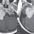
Anomalies of the semicircular canals (SCCs) may coincide with the presence of a morphologically normal cochlea. These anomalies are often associated with other anomalies of the inner ear; thus they should be considered as part of diffuse inner ear malformations. SCC anomalies are often bilateral, with one side is more affected than the other. There is a high association between SCC aplasia and the CHARGE association (coloboma of the eye, congenital heart defects, choanal atresia, developmental and growth retardation, genital hypoplasia, and ear anomalies/or deafness). Isolated aplasia of the posterior semicircular duct has been described in patients with Wartenberg and Alagille syndrome.
Clinical Features
The most common presenting symptom is sensorineural hearing loss, even when the cochlea is normal on imaging. Conductive hearing loss is often present due to associated oval window atresia and ossicular chain malformation. Dizziness is present in only 19% of the patients. A few patients complain of Tullio phenomenon, which is dizziness upon hearing loud noises.
Pathology
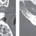
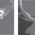
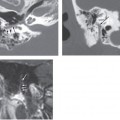
The vestibule is the largest cavity of the bony labyrinth. The saccule and utricle reside within the vestibule, with the saccule located anteroinferiorly anchored to the spherical recess, and the utricle located posterosuperiorly anchored to the elliptical recess. The utriculosaccular duct connects the saccule and the utricle together. On the posterior wall of the utricle reside five openings for the three semicircular ducts (the superior and posterior canals have a common crus). Each of the SCCs is an arc of approximately two thirds of a full circle, oriented at right angles to each other. The superior SCC is in an oblique sagittal plane, approximately perpendicular to the long axis of the petrous bone. The vestibule is considered abnormally large when the ratio of the transverse diameter of the vestibule in the axial plane to the inner diameter of the lateral semicircular canal exceeds 1:2.
The development of the semicircular canals is completed between the 19th and 22nd week of gestation. Its development starts at the 4th week, along with the other inner ear structures, from the vestibular anlage. It begins as two fingerlike extensions from the utricular side of the otic vesicle. Between weeks 4 and 8, the membranous labyrinth has three subdivisions: the saccule with the cochlear duct (pars inferior), the utricle with its SCCs (pars superior), and the endolymphatic duct system. Embryologically, the pars superior, semicircular canals, and utricle are phylogenetically older than the pars inferior, cochlea, and saccule. As a result, developmental malformations are more common in the neolabyrinth (cochlea and saccule) than in the paleolabyrinth (SCCs and utricle). Complete maturation of the SCC occurs at about mid-gestation, with the superior SCC completed first and the lateral SCC completed last.
Related posts:

Stay updated, free articles. Join our Telegram channel

Full access? Get Clinical Tree


