12 Shoulder Examination
♦ Setup
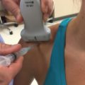
• The patient should be seated on a rotating stool facing the ultrasound screen.
• The examiner stands behind the patient so that both the patient and examiner can see the screen.
♦ Normal Anatomy
Long Head of the Biceps and Subscapularis
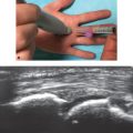
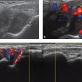
• The probe should be placed parallel to floor, centered over the anterior aspect of the shoulder (Fig. 12.1).
• The elbow should be flexed to 90 degrees; rotation of the shoulder is neutral.
• The long head of the biceps should be located and traced distally to the myotendinous junction. The pectoralis major tendon inserts just lateral to the biceps tendon at this point.

Fig. 12.1 The long head of the biceps viewed in the short axis.
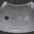
• The probe should be moved proximal and medial; the subscapularis can then be visualized on its long axis (Fig. 12.2).
• The patient should slowly externally rotate to confirm that the biceps is not subluxating medially out of the groove.
• The subscapularis should be scanned both proximally and distally.
• The coracoid process is visible medial to the subscapularis tendon.
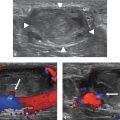
• The probe should then be turned 90 degrees to visualize the long head of the biceps along its long axis between the greater and lesser tuberosities (Fig. 12.3).
• The probe is moved medially to visualize the subscapularis along the short axis (Fig. 12.4).

Fig. 12.2 The subscapularis viewed in the long axis.

Fig. 12.3 The long head of the biceps viewed in the long axis.

Tips
• When first learning ultrasound of the shoulder, it is easiest to locate bony landmarks first (greater and lesser tuberosities, humeral head, and glenoid), then begin identifying soft tissue structures.
• To quickly locate the biceps in the long axis, the operator should scan medially and laterally with the probe oriented vertically; the biceps will be located between the lesser and greater tuberosities.
• When visualizing the long head of the biceps in the longitudinal axis, the probe often must be oriented in a proximal lateral-distal-medial direction to capture the entire tendon in the plane, because the tendon takes a lateral course before entering the glenohumeral joint.
• The location of the biceps tendon should be noted. Subluxation deep to the subscapularis indicates a subscapularis tear; subluxation superficial to the tendon indicates a medial biceps pulley lesion.
• Any fluid around the biceps tendon is indicative of pathology. When fluid is noted both in the bicipital sheath and superficial to the subscapularis, a full-thickness rotator cuff tear is highly likely.
Supraspinatus and Infraspinatus
• The patient should put the opposite hand on the ipsilateral iliac crest and adduct the elbow.
• The probe should be placed on the most anterior aspect of the shoulder oriented toward the ipsilateral hip. This provides a view of the anterior supraspinatus in the long axis.
• The probe is moved anteriorly until the biceps tendon is visualized as it crosses into the glenohumeral joint (Fig. 12.5). When the biceps tendon is visualized, the probe should be slowly moved posteriorly.
• As the probe is moved posteriorly, the posterior fibers of the supraspinatus (Fig. 12.6a,b) and the infraspinatus will be visualized (Fig. 12.6c,d).