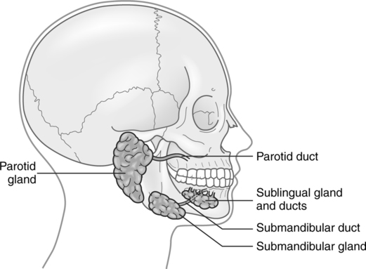
Sialography

CHAPTER 22


Sialography


The radiographic visualization of the salivary glands and ducts is called sialography. The evaluation of the salivary glands is most often accomplished with computed tomography or magnetic resonance imaging; however, sialography becomes the method of choice when a definitive diagnosis is required for pathology such as sialadenitis (inflammation of the salivary glands) and the oral component of ’Sjögren’s syndrome (an autoimmune disease process that causes dry eyes and dry mouth). Sialography involves the introduction of a water-soluble contrast agent into the orifices of the salivary ducts. In most cases, this procedure requires a minimum of specialized equipment and can be performed in a regular radiographic or fluoroscopic room.


ANATOMIC CONSIDERATIONS


The salivary glands secrete saliva into the mouth. Saliva is a liquid that is approximately 99% water. The salivary glands secrete between 1000 and 2000 ml of saliva every day. Found in the saliva are basically two types of materials secreted by the salivary glands—mucus, a combination of mucin and water that is a very viscous substance used to lubricate food particles and maintain oral hygiene, and serous fluid, which contains the enzyme amylase, a substance that begins the digestive process of starches.


There are three pairs of salivary glands—the parotid glands, submandibular glands, and sublingual glands (Fig. 22-1).


PROCEDURE


Scout films of the area of interest may or may not be taken. In cases of suspected sialolithiasis (salivary gland or salivary duct calculi), scout films are mandatory. In cases in which scout films are not taken, stones may be obliterated by the contrast medium, causing an inaccurate diagnosis to be made. The study may be performed with fluoroscopic visualization and spot filming as well as with overhead radiographic projections. Computed tomography, ultrasonography, and magnetic resonance imaging are replacing conventional sialography. Although the primary means of diagnosis has been ultrasonography, conventional sialography still has a place in the realm of interventional radiography for the treatment of pathology of the salivary glands.


After scout films have been taken, the radiologist locates the orifices of the salivary ducts by having the patient express some saliva. The physician can either palpate the salivary gland or have the patient suck on a lemon slice. When the salivary duct is located, it is dilated with standard double-ended blunt dilators or with silver lacrimal probes. After dilation of the orifice, the duct is cannulated. Several types of cannulas are available, but the use of a modified Abbott butterfly set has proved successful in most cases, especially if the patient has to be moved to another location for additional study. The modification is accomplished by filing the beveled tip of the needle flat and smooth with a medium-fine metal file. The wings of the butterfly are then secured with a hemostat. This allows for ease of insertion of the cannula into the duct. The cannula should be prefilled with the contrast medium to avoid injection of air bubbles.

Stay updated, free articles. Join our Telegram channel

Feb 27, 2016 | Posted by in GENERAL RADIOLOGY | Comments Off on Sialography

Full access? Get Clinical Tree

Get Clinical Tree app for offline access