Simple Congenital Heart Disease


  • The distinction between “simple” and “complex” congenital heart disease is not well defined. In this chapter, simple congenital heart disease describes uncomplicated anatomic defects or shunt lesions that are not associated with other cardiovascular anomalies.


  • This chapter describes cardiovascular magnetic resonance (CMR) findings in the following congenital cardiac anomalies:


    • Interatrial communications


    • Ventricular septal defect


    • Patent ductus arteriosus


    • Partially anomalous pulmonary venous connection


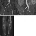
    • Coarctation of the aorta and bicommissural aortic valve


  • CMR is usually used in concert with other imaging modalities for assessment of cardiovascular anatomy and function, measurements of blood flow, tissue characterization, and for evaluation of myocardial perfusion and viability.


  • CMR is particularly helpful in adolescent and adult patients with congenital heart disease because it overcomes many of the limitations associated with echocardiography (e.g., restricted acoustic windows), cardiac catheterization (e.g., invasive, expensive, radiation exposure), computed tomography (e.g., radiation exposure, predominantly static anatomic information), and nuclear scintigraphy (e.g., low spatial resolution, lack of anatomic information, radiation exposure).


KEY POINTS


INTERATRIAL COMMUNCATIONS


Types of Interatrial Communications


Types of interatrial communications are shown in Figure 14-1 . Patent foramen ovale (PFO) is the most common type of interatrial communication. Anatomically, it is located between normally formed septum primum and septum secundum. The second most common type of interatrial communication is secundum atrial septal defect, followed by primum atrial septal defect.


Figure 14-1


Types of interatrial communications. A, Cross-section of the atria demonstrating the atrial septal components. Patent foramen ovale (PFO) is the most common type of interatrial communication. Anatomically, it is located between normally formed septum primum and septum secundum ( arrow ). AVS, atrioventricular septum; FO, fossa ovalis; ILB, inferior limbic band; LA, left atrium; LV, left ventricle; RA, right atrium; RV, right ventricle; Sept. 1 0 , septum primum; SLB, superior limbic band (septum secundum). B, The second most common type of interatrial communication is secundum atrial septal defect (ASD 2 0 ), followed by primum atrial septal defect (ASD 1 0 ). ASD 1 0 , primum atrial septal defect; ASD 2 0 , secundum atrial septal defect.


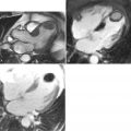
Secundum atrial septal defect ( Figures 14-2 and 14-3 ) is a defect within the fossa ovalis, usually caused by a single or multiple defects within septum primum. Septum secundum is usually well formed. Most secundum atrial septal defects (ASDs) are not confluent with the vena cavae, right pulmonary veins, coronary sinus, or the atrioventricular (AV) valves. Primum atrial septal defect ( Figures 14-4 to 14-6 ) is an endocardial cushion (AV canal) defect with an interatrial communication located between the anterior-inferior margin of the fossa ovalis and the AV valves. It is considered a form of partial AV canal defect with two separate AV valve annuli and no ventricular septal defect of the AV canal type. Sinus venosus defect is a communication between one or more of the right pulmonary veins and the cardiac end of the superior vena cava (SVC) and/or the posterior wall of the right atrium. Sinus venosus defect ( Figures 14-7 to 14-9 ) comprises approximately 4% to 11% of interatrial communications. Although anatomically not a true atrial septal defect, sinus venosus defect results in an interatrial communication and is hemodynamically similar to an atrial septal defect.


Figure 14-2


ECG-gated steady-state free precession (SSFP) cine magnetic resonance (MR) image in the four-chamber plane showing a large secundum atrial septal defect. The defect results from a deficiency of septum primum. The hemodynamic consequence is a left-to-right shunt across the fossa ovalis. Note the dilated right atrium (RA) and right ventricle (RV). By velocity encoded cine MR measurements in the main pulmonary artery and ascending aorta, the pulmonary-to-systemic flow ratio measured 1.6. The defect was subsequently closed in the cardiac catheterization laboratory with an Amplatzer septal occluder.


Figure 14-3


Secundum atrial septal defect. Left panel: ECG-gated SSFP cine MR image in an oblique sagittal plane of the atrial septum, demonstrating a small secundum atrial septal defect. Right panel: ECG-gated velocity encoded cine MR in the same plane. Flow velocity is encoded in the anterior-posterior direction (in-plane; see arrow above color scale). Flow velocity and direction are color-encoded. Blue indicates flow from posterior (left atrium; LA) to anterior (right atrium; RA). Note the left-to-right flow jet through the secundum atrial septal defect.


Figure 14-4


ECG-gated SSFP cine MR image in the axial plane in an infant with heterotaxy syndrome and polysplenia. Note the dextrocardia and the large primum atrial septal defect ( arrow ). CMR was performed to evaluate the anatomy of the pulmonary veins to exclude stenosis as a potential cause of severe pulmonary hypertension. Note two unobstructed pulmonary venous connections into the left atrium. LA, left atrium; LV, left ventricle; RA, right atrium; RV, right ventricle; LLPV, left lower pulmonary vein; RLPV, right lower pulmonary vein.


Figure 14-5


CMR imaging of primum atrial septal defect. Left panel: ECG-gated, breath-hold cine SSFP MR image in the four-chamber plane. The primum atrial septal defect is located between the inferior rim of the fossa ovalis and the attachments of the AV valves to the ventricular septal crest ( arrow ). Note the intact fossa ovalis. Right panel: ECG-gated velocity encoded cine MR in the same plane. Flow velocity is encoded in the anterior-posterior direction (in-plane; see arrow next to color scale). Flow velocity and direction are color-encoded. Blue indicates flow from posterior (left atrium; LA) to anterior (right atrium; RA) through the primum atrial septal defect. Note the absence of flow at the ventricular level indicating absence of a ventricular septal defect component. By velocity encoded cine MR measurements in the main pulmonary artery and ascending aorta, the pulmonary-to-systemic flow ratio measured 1.7. The primum atrial septal defect was subsequently surgically closed with a pericardial patch.


Figure 14-6


Cleft anterior mitral valve leaflet in a patient with primum atrial septal defect. The anatomy of the AV valves is clearly seen on ECG-gated breath-hold cine SSFP MR images acquired in the short axis plane at the base of the ventricles. A, Diastolic frame showing the cleft between the superior and inferior components of the anterior mitral leaflet. The cleft is pointing toward the ventricular septal crest ( arrow ). B, Systolic frame showing the plane of coaptation between the superior and inferior components of the anterior mitral leaflet. IC, inferior component of the anterior mitral valve leaflet; SC, superior component of the anterior mitral valve leaflet; ML, mural (posterior) mitral valve leaflet.


Figure 14-7


Superior vena cava (SVC)-type sinus venosus defect. A, Axial plane ECG-gated breath-hold cine SSFP MR image depicting a defect between the posterior wall of the superior vena cava (SVC) and the anterior wall of the right pulmonary vein (RPV) ( arrow ). The communication between the left atrium (LA) and the superior vena cava is through the left atrial termination of the right pulmonary vein (*). B, The sinus venosus septum is depicted by the dashed line. Note the “normal” drainage of the right pulmonary vein once the sinus venosus septum is “restored.” Ao, aorta.


Figure 14-8


Superior vena cava (SVC)-type sinus venosus defect. A, ECG-gated breath-hold cine SSFP MR images acquired in an oblique sagittal plane, demonstrating a large defect between the posterior wall of the superior vena cava (SVC) and the anterior wall of the right upper pulmonary vein (*). Note that the resultant defect allows a communication between the left atrium (LA) and the superior vena cava. Therefore this defect is not considered an atrial septal defect (ASD) because there is no direct communication between the left and right atria. B and C, Systolic and diastolic frames from ECG-gated velocity encoded cine MR in the same plane as A . Flow velocity is encoded in the superior-inferior direction (in-plane; see arrow next to color scale in panel C ). Flow velocity and direction are color encoded. Blue indicates flow from superior to inferior and red indicates flow from inferior to superior. Note the bidirectional flow through the sinus venosus defect. The net pulmonary-to-systemic flow ratio, evaluated by through-plane velocity encoded cine MR perpendicular to the ascending aorta and main pulmonary artery, measured 2.3.


Figure 14-9


Right ventricular volume load in a patient with SVC-type sinus venosus defect and a large left-to-right shunt with pulmonary-to-systemic flow ratio of 2.3. Ventricular short-axis ECG-gated breath-hold SSFP cine MR images in diastole ( A ) and systole ( B ). Note the dilated right ventricle and the flat systolic configuration of the interventricular septum in diastole ( A ) indicating volume overload. In systole ( B ), the interventricular septum maintains its flat configuration, indicating right ventricular hypertension. This patient was treated for pulmonary hypertension prior to successful surgical closure of her sinus venosus defect.


Measurement of Pulmonary-to-Systemic Flow Ratio in Patients with Interatrial Communications


In the absence of associated cardiovascular anomalies and with normal pulmonary vascular resistance, interatrial communications result in a left-to-right shunt proximal to the AV valves. The volume of the left-to-right shunt depends on the size of the defect and the relative compliance of the right and left ventricles. Most patients are asymptomatic during childhood and adolescence. Some patients with unrepaired interatrial communications will develop complications during adult life, including pulmonary vascular disease, atrial arrhythmias, heart failure, and paradoxical emboli. Indications for closure of interatrial communications in children include symptoms (e.g., exercise intolerance, dyspnea, slow weight gain), left-to-right shunt greater than 1.5 to 2.0 associated with right ventricular volume overload, and paradoxical emboli. Although a patent foramen ovale or a small secundum atrial septal defect may decrease in size or even close spontaneously during infancy and early childhood, larger secundum atrial septal defects can increase in size during adulthood. Primum atrial septal defects and sinus venosus defects almost never decrease in size over time. CMR allows evaluation of the location and size of the interatrial communication as well as quantitative assessment of the hemodynamic burden from the defect(s), such as pulmonary-to-systemic flow ratio, right ventricular size and function, etc. ( Figures 14-10 and 14-11 ).


Figure 14-10


ECG-gated, breathe-through velocity encoded cine MR perpendicular to the ascending aorta ( A ) and main pulmonary artery ( B ). C, Graph showing flow rate (Y axis) versus time (X axis) during the cardiac cycle. Stroke volume is the area under the flow rate-time curve. Multiplying the stroke volume by the heart rate yields the flow per minute (L/min) through the blood vessel. In this example, flow through the ascending aorta equals systemic blood flow (Qs) and flow through the main pulmonary artery equals pulmonary blood flow (Qp).


Figure 14-11


ECG-gated, breathe-through velocity encoded cine MR perpendicular to the mitral ( red contour ) and tricuspid ( green contour ) valves ( A ). B, Graph showing flow rate (Y axis) versus time (X axis) during diastole. Stroke volume is the area under the flow rate-time curve. Multiplying the stroke volume by the heart rate yields the flow per minute (L/min). In patients with interatrial communications, flow through the mitral valve (MV) equals systemic flow (Qs) and flow through the tricuspid valve (TV) equals pulmonary blood flow (Qp). See Figure 14-19 for other examples of shunt calculations.


VENTRICULAR SEPTAL DEFECTS


Different types of ventricular septal defects (VSDs) are shown in Figure 14-12 .


Figure 14-12


Schematic diagram illustrating different types of ventricular septal defects (VSDs).


Conoventricular septal defect ( Figure 14-13 ) results from malalignment between the conal (outlet) septum superiorly and the muscular ventricular septum inferiorly. A conoventricular septal defect may be membranous if its posterior-inferior border is confluent with the tricuspid valve or may be muscular if the inferior limb of septal band forms its inferior-posterior border.


Figure 14-13


ECG-gated breath-hold SSFP cine MR images showing a conoventricular VSD in a 49-year-old woman with pulmonary vascular disease and cyanosis. A, Ventricular short-axis diastolic image demonstrating the defect between the conal septum superiorly ( white arrow ) and muscular interventricular septum inferiorly ( black arrow ). Note mild anterior malalignment of the conal septum and an unobstructed infundibulum (Inf). B, Four-chamber view showing the subaortic (*) location of the conoventricular septal defect ( arrow ). Pulmonary-to-systemic flow ratio measured 0.68. LA, left atrium; LV, left ventricle; RA, right atrium; RV, right ventricle.

Only gold members can continue reading. Log In or Register to continue

Stay updated, free articles. Join our Telegram channel

Feb 1, 2019 | Posted by in MAGNETIC RESONANCE IMAGING | Comments Off on Simple Congenital Heart Disease

Full access? Get Clinical Tree

Get Clinical Tree app for offline access