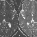
107 SNR at 3 T
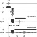
The signal intensity of each voxel displayed in MRI depends upon multiple tissue characteristics, two of these being T1 and T2, as well as the parameters specified for the acquired pulse sequence, for example TR, TE, TI, and flip angle. However, there are many additional factors that also influence signal intensity. As mentioned in previous cases, the signal intensity of each voxel is directly proportional to the number of mobile protons within the volume element. Increasing the voxel size thus increases SNR, however this decreases spatial resolution-an undesirable effect in clinical imaging. This means that the higher the resolution, the lower the SNR of the underlying displayed volume element. A further factor influencing SNR is the field strength used for MR imaging, with the desire for higher SNR principally driving the move to ever-higher clinical field strengths.
 Field Strength
With increasing magnetic field strength, the energy difference between the two levels that protons (spins) can occupy is increased. As a consequence, a greater number of spins occupy the lower energy level prior to any excitation. Thus, with increasing field strength, more spins are available to be excited to the higher energy level, together with more energy needed to excite a spin from the lower to the higher level, resulting in a stronger signal being emitted when the spins return after excitation to the lower energy level. This gain in SNR, according to theory, is directly proportional to the increase in field strength.
Related posts:
Stay updated, free articles. Join our Telegram channel

Full access? Get Clinical Tree


