and Zdeněk Fryšák1
(1)
Department of Internal Medicine III – Nephrology, Rheumatology and Endocrinology, Faculty of Medicine and Dentistry, Palacky University Olomouc and University Hospital Olomouc, Olomouc, Czech Republic
Keywords
Solid noduleHalo sign8.1 Essential Facts
According to The 2015 American Thyroid Association (ATA) Guidelines: isoechoic or hyperechoic solid nodule, or partially cystic nodule with eccentric uniformly solid areas without microcalcifications, irregular margin or extrathyroidal extension, or taller than wide shape prompts low suspicion for malignancy 5–10% [1].
Caution! About 15–20% of thyroid cancers are isoechoic or hyperechoic on US, and these are generally the follicular variant of papillary thyroid carcinoma (PTC) or follicular thyroid carcinoma (FTC) [1].
8.2 US Features of a Benign Solid Nodule
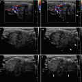
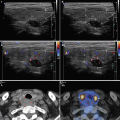
US findings for benign nodule (Fig. 8.1aa) [2]:


Fig. 8.1
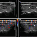
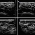
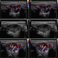
(aa) A 24-year-old woman with solitary solid nodule (arrowheads), size 16 × 14 × 9 mm and volume 1 mL in the RL. According to US criteria typically non-suspicious, benign nodule: ovoid shape, “more wide than tall”; homogeneous structure; isoechoic; well-defined with thin halo sign; Tvol 16 mL, RL 9 mL, and LL 7 mL; transverse. (bb) Detail of solitary solid nodule (arrowheads): ovoid shape; homogeneous structure; isoechoic; well-defined with thin halo sign. (cc) Detail of solitary solid nodule (arrowheads), CFDS: increased vascularity at periphery and sporadic parenchymal vascularity, pattern I; transverse. (dd) Detail of solitary solid nodule (arrowheads): ovoid shape; homogeneous structure; isoechoic; well-defined with thin halo sign; longitudinal. (ee) Detail of solitary solid nodule (arrowheads), CFDS: peripheral vascularity and one intranodal vessel branch, pattern I; longitudinal


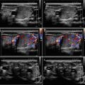
Fig. 8.2
(aa) A 57-year-old woman with solitary solid nodule (arrowheads), size 14 × 13 × 6 mm and volume 0.6 mL in the right branch of isthmus. According to US criteria typically non-suspicious, benign nodule: elliptical shape, “more wide than tall”; homogeneous structure; isoechoic; well-defined with thin halo sign; Tvol 19 mL, RL 9 mL, and LL 10 mL; transverse. (bb) Detail of isthmic solitary solid nodule (arrowheads), CFDS: peripheral vascularity and minimal parenchymal vascularity, pattern I; transverse. (cc) Detail of isthmic solitary solid nodule (arrowheads): ovoid shape; homogeneous structure; isoechoic; well-defined with thin halo sign; slightly enlarged “mirror artifact” (arrows) of the nodule visible into the trachea; longitudinal. (dd) Detail of isthmic solitary solid nodule (arrowheads), CFDS: increased peripheral vascularity, pattern I; slightly enlarged avascular “mirror artifact” (arrows) of the nodule, visible into the trachea; longitudinal
Stay updated, free articles. Join our Telegram channel

Full access? Get Clinical Tree


