Solutions to Test Yourself!
The exercises and solutions have been numbered consecutively. Some of the exercises have several different correct solutions. If the exercises can be solved simply by referring to the chapters in the book, I have indicated where you will find the necessary information.
After you have completed the exercises, compare your score and results with those of your colleagues. The score on the right gives you an impression of the degree of difficulty. Enjoy the challenge!
Solution to exercise 1 (p. 32):
9 Points
You will find the sequence for interpreting CCTs on page 26. Each step gives you 1/2 point with 3 extra points for the correct sequence, which adds up to 9.
Solution to exercise 2 (p. 45):
9 Points
Solution to exercise 3 (p. 45):
10 Points
Solution to exercise 4 (p. 45):
6 Points
Solution to exercise 5 (p. 45):
2 Points
Tubular and nodular structures can be differentiated by comparing a series of images.
Solution to exercise 6 (p. 45):
3 Points
Vessels in which beam-hardening artifacts occur because of CM inflow are the superior vena cava, inferior vena cava, and the subclavian vein.
Solution to exercise 7 (p. 48):
3 Points
Fractures, inflammatory processes, and tumors or metastases can cause swelling of mucous membranes and retention of fluids in the mastoid sinuses and middle ear; these are normally filled with air.
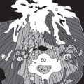
Solution to exercise 8 (p. 57):
18 Points
This image requires careful study. You will discover several types of intracranial hemorrhage and the complications resulting from them.
Solution to exercise 9 (p. 72):
9 Points
Gray and white matter appear well defined on narrow brain windows.
Level | Width | Gray scale |
+ 35 HU | 80 HU | – 5 HU to + 75 HU |
3
CCT sections are normally oriented parallel to the orbitomeatal line, |
|
so that initial and follow-up studies can be precisely compared. | 2 |
2-mm sections at 4-mm increments are acquired through the petrosal bone, | 2 |
then thickness and table movement are set at 8 mm. | 2 |
Solution to exercise 10 (p. 72):
16 Points
Solution to exercise 11 (p. 72):
2 Points
Subarachnoid hemorrhage in children may be visible only next to the falx or in the lateral (Sylvian) fissure.
Solution to exercise 12 (p. 72):
10 Points
Practice makes perfect!
Solution to exercise 13 (p. 72):
4 Points
Fracture of the right frontal bone and absent right frontal sinus (the latter is a congenital variation, not a hemorrhage, as indicated by the osseous trabeculae)
Solution to exercise 14 (p. 72):
8 Points
This was a difficult question. In the left internal jugular vein there is unusual sedimentation of the CM due to slow blood flow. The asymmetry of the jugular veins is not a sign of thrombosis. A left cervical abscess makes the neck muscles appear poorly defined.
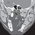
Solution to exercise 15 (p. 73):
4 Points
In this patient the surface subarachnoid spaces are clearly too narrow and the ventricles distended. These signs indicate that CSF drainage is reduced or blocked and there is imminent danger of brain herniation. There is generalized brain edema. A neurosurgeon should be consulted about inserting an intraventricular shunt.
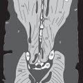
Solution to exercise 16 (p. 73):
3 Points
It is possible to mistake the subarachnoid hemorrhage around the left frontal lobe as an artifact. The left frontal cortex is outlined by blood. If you did not see any abnormality, return to the chapter about the head.
Solution to exercise 17 (p. 73):
You have of course taken the hint about not giving up too soon; the right medial rectus muscle (47c) is thickened. It is the second muscle to become involved in endocrine ophthalmopathy.
If you cannot remember which muscle is affected first, return to page 61.
Related posts:


Stay updated, free articles. Join our Telegram channel

Full access? Get Clinical Tree


