Alan D. Birney • Gary P. Siskin
Since 1974, polyvinyl alcohol (PVA) particles have been used as a particulate agent for embolization procedures.1 However, as experience was gained with this agent, its inherent limitations and disadvantages were recognized. These include size variability in a given preparation of particles due to the manufacturing process, particle aggregation, and microcatheter occlusion during delivery. Spherical embolic agents were developed in response to these limitations and become increasingly popular since their introduction.2
DEVICE DESCRIPTION
Trisacryl Gelatin Microspheres
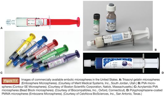
In 1996, Laurent et al.3 reported on the development of a spherical, nonresorbable embolization agent. These microspheres (Embosphere Microspheres; Merit Medical Systems, Inc., South Jordan, Utah) consist of a trisacryl polymer that is impregnated and embedded with gelatin (Fig. 7.1A). Trisacryl gelatin microspheres had been initially manufactured in the mid-1980s for use as a microcarrier for cell cultures.4 These microspheres are biocompatible, hydrophilic, and deformable. In addition, cellular adhesion to these microspheres is supported by the presence of denatured collagen on their surface.3,4
Interventionalists instantly accepted these microspheres once they became commercially available because they successfully addressed the limitations of particulate PVA. The manufacturing process of these microspheres enabled a more narrow and reliable range of particles to be produced.2,3 In addition, these microspheres did not aggregate, possibly due to their spherical configuration, the presence of a positive surface charge on the microspheres, and their hydrophilic nature.2,5,6 In addition, a more predictable target occlusion could be achieved due to the fact that these microspheres did not aggregate. Derdeyn et al.5 demonstrated that trisacryl gelatin microspheres occlude more distally than PVA particles of matched size, supporting the absence of aggregation when these microspheres are used. In fact, the level of vascular occlusion appears to correlate closely with the diameter of the microspheres used for embolization.6–8 The narrow size range of microspheres, their lack of aggregation, and their deformability minimize the risk of microcatheter occlusion and contribute to the ease of delivery during administration.
The cellular response to embolization with trisacryl gelatin microspheres has been described. Macrophages, polymorphonuclear cells, and sparse lymphocytes have all been described after embolization with these microspheres, as has vessel recanalization.3,9–11 Interestingly, these microspheres often undergo transvascular migration and can be found within the vessel lumen, within the vessel wall, or completely outside of the vessel after embolization11; PVA particles are less likely to be found outside of the vessel. This has been attributed to the inflammatory reaction induced by these microspheres.
Polyvinyl Alcohol Microspheres
Given the decades of success seen with embolization procedures performed with particulate PVA, it seems logical that a PVA-based microsphere (Contour SE Microspheres; Boston Scientific Corporation, Natick, Massachusetts) would be developed as a next generation embolic agent (Fig. 7.1B). PVA microspheres appear to generate a milder inflammatory response than both particulate PVA and trisacryl gelatin microspheres.9 After embolization, neutrophils are acutely seen, but these are ultimately replaced by macrophages and occasional lymphocytes.
PVA microspheres, like trisacryl gelatin microspheres, address the disadvantages of particulate PVA with better size uniformity, less aggregation, and ease of administration. However, many of the inherent properties differ between these microspheres, including compressibility and elastic recovery.10 PVA microspheres have been shown to be highly compressible with a delayed and incomplete elastic recovery, leading to a more distal embolization when compared to other spherical embolic agents.7,8 This is felt to be due to change in shape of the particle with compression during delivery, which allows them to deform and occlude more distally than intended.
Acrylamido PVA microspheres (Bead Block microspheres; Biocompatibles, Inc., Oxford, Connecticut) consist of a PVA-based hydrogel polymer (Fig. 7.1C). Its properties have been shown to be intermediate between spherical PVA microspheres and trisacryl gelatin microspheres, with similar compression and nearly immediate subsequent reexpansion when compared with trisacryl gelatin microspheres.2 These microspheres have been found to occlude slightly more distal than trisacryl gelatin microspheres, which is likely due to the slight differences in force required to compress the microspheres.
Polyphosphazene-Coated PMMA Microspheres
Polyphosphazene-coated polymethylmethacrylate (PMMA) microspheres (Embozene Microspheres; CeloNova BioSciences, Inc., San Antonio, Texas) consist of a Polyzene-F shell surrounding a hydrogel core of PMMA (Fig. 7.1D). When PMMA undergoes an alkaline hydrolysis, its structural flexibility increase, making it an appropriate material to use for an embolic agent.12 This was theorized as early as 1989 by Jayakrishnan et al.13 Polyzene-F is a proprietary, biocompatible, and nonresorbable version of the poly(bis[trifluoroethoxy]phosphazene) (PTFEP) polymer class and is applied as a thin coating on the PMMA core.14 Polyzene-F has previously been used in vascular stents where it was found to have an absence of a significant inflammatory reaction.15 A preclinical evaluation by Stampfl et al.14 demonstrated similar findings, with only a minimal lymphocyte-mediated inflammatory reaction noted after embolization in a renal artery model. This is different from the findings of Verret et al.,12 which noted early recruitment of phagocytic cells in a uterine artery model. In terms of the distribution of these microspheres within embolized vasculature, Verret et al.16 demonstrated that they occlude more distal vessels than similarly sized trisacryl gelatin microspheres. They attributed this finding to their compressibility and deformability, enabling them to conclude that deformability determines the size of the vessel occluded as opposed to the actual particle size, which potentially makes the level of occlusion unpredictable with these microspheres.
TECHNIQUE
In general, spherical microspheres are packaged in a syringe consisting of a defined volume of embolic and normal saline. For administration, contrast material is added to make the solution radiopaque. A maximum saline-to-contrast ratio of 1:1 is recommended. Typically, gentle swirling of the solution is recommended before delivery; this and time will allow for the microspheres to be adequately suspended in the saline and contrast solution. To-and-fro aspiration between two syringes connected via a three-way stopcock is both unnecessary and not recommended as it may damage the microspheres.
Once the microspheres have been prepared, they are typically administered through a microcatheter under fluoroscopic guidance. Although the catheter selection can be based on the size of the microspheres being used for any given procedure, these microspheres tend to be more easily administered with microcatheters having an inner diameter of 0.027 to 0.028 in as opposed to smaller microcatheters.
One limitation of the trisacryl gelatin microspheres is that they are clear, which can make preparation difficult and visual confirmation of delivery from the syringe challenging. A subsequent product used elemental gold to stain the microspheres, which improved visualization. However, the addition of elemental gold resulted in a greater degree of inflammation after embolization, which ultimately led to the discontinuation of this product.17
CLINICAL APPLICATIONS
Beaujeux et al.10 reported the initial clinical success of using trisacryl gelatin microspheres on 105 patients with tumors or arteriovenous malformations in the head, neck, or spine. Since this initial report, all of the available embolic microspheres have been rapidly incorporated into present-day embolization procedures and have been used successfully for various indications that are suitable for a particulate embolic agent.
These microspheres have had particularly notable success when used for uterine artery embolization (UAE) to treat symptomatic uterine fibroids. Although the success seen with the use of trisacryl gelatin microspheres has set the standard for subsequent comparative clinical trials,18 each of the available embolic microspheres have demonstrated high clinical success and fibroid infarction rates in association with this procedure.19,20 However, all of the attention paid to this procedure has brought to light how the different characteristics of these embolic microspheres can affect clinical outcomes and as a result has improved our understanding of these agents. For example, Contour SE Microspheres have been shown to be less effective for this procedure than other agents.21,22 This is felt to be due to the differences in compressibility, deformability, and elasticity of this particular product when compared to the other available embolic microspheres, which was previously not appreciated until the differences in clinical outcomes were recognized.
NEXT GENERATION MICROSPHERES
Resorbable Microspheres
In theory, there is an inherent appeal to the concept of resorbable microspheres. All embolization procedures are being performed for certain indications, and if the goal of the procedure can be accomplished without implanting a permanent foreign body into the patient, then why would not that be preferred? In 2007, Laurent2 suggested that the ideal resorbable microsphere would have four characteristics: it would have controlled resorption time, it would cause only a limited local inflammatory response, it would be associated with a complete functionality of the target vascular bed, and it would be loadable. If these expectations could be met, then one could be assured that the target pathology would be definitively and appropriately treated while leaving open the possibility that the target organ would fully recover and suffer no long-term effects from the embolization procedure.
There has been significant effort to develop bioresorbable microspheres in recent years. Nowadays, degradable starch microspheres have been evaluated for several years23 and are commercially available (EmboCept S; PharmaCept, Berlin, Germany). These microspheres have been used for liver-directed tumor therapy.24,25 The problem is that the starch microspheres are only available in a 50-μm diameter size and have a very short half-life, limiting their acceptance for the more common applications requiring particulate embolization.
Weng et al.26 have done much work with bioresorbable hydrogel microspheres prepared from carboxymethylcellulose and chitosan. Chitosan (N-acetylglucosamine) is a linear polysaccharide derived from chitin after deacetylation.27 Introducing carboxymethyl groups into the chitosan produces chitosan derivatives that are readily soluble in a physiologic pH.28 The carboxymethyl chitosan can then be cross-linked with oxidized carboxymethylcellulose to form microspheres through an inverse emulsion method. These microspheres are degraded by lysozyme into glucosamine, which can be absorbed completely by the body. The degradation time appears to depend on the parameters of microsphere preparation, specifically on the degree of cross-linking density. These microspheres do not aggregate due to low coefficient of surface friction. In vivo evaluation comparing these microspheres with trisacryl gelatin microspheres has been performed with renal embolization in a rabbit model.29 The performance of the two agents was similar in terms of the mean size of the vessels occluded with the two microspheres. These microspheres were biocompatible and well tolerated, with a mild tissue reaction and no evidence of vessel wall disruption. The authors did demonstrate that preparing the resorbable microspheres with a higher cross-linking density makes the spheres more rigid, which subsequently causes a more proximal embolization. Of note, the carboxylic groups in the microsphere matrix and their highly porous internal structure may allow for loading and release of positively charged drugs such as doxorubicin hydrochloride.28
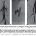
Microspheres consisting of poly(lactic-co-glycolic acid) (PLGA) coated with type I bovine fibrillar collagen have also been evaluated (Occlusin 500 Artificial Embolization Device; IMBiotechnologies Ltd., Edmonton, Alberta, Canada). These noncompressible microspheres create a vascular occlusion mechanically and by binding platelets. An in vivo evaluation of these microspheres has been performed with UAE in a sheep model.30 In this study, the effects of embolization with 150- to 212-μm PLGA microspheres were compared with the effects after embolization with trisacryl gelatin microspheres. At 6 months, none of the embolic material in the animals embolized with PLGA microspheres was detectable. At 12 months, all uterine arteries treated with the PLGA microspheres were recanalized or in the process of being recanalized; recanalized vessels were histologically indistinguishable from untreated vessels. The arteries treated with trisacryl gelatin microspheres remained occluded at 12 months.
There are other agents in various stages of development. Water-soluble PVA microspheres have been developed and evaluated in a pig kidney model.31 The duration of arterial occlusion using these microspheres is possibly related to the degree of saponification of PVA. Microspheres manufactured from hydrophilic methacrylate monomer copolymerized with degradable cross-linkers have been evaluated as well.32
Visible Microspheres
An additional point of development for embolic microspheres is in the area of radiographic visibility. Nowadays, microspheres used for embolization are not radiographically visible and must therefore be mixed with contrast to be seen under fluoroscopy during delivery. An agent that could be visualized under fluoroscopy as well as under computed tomography (CT) or magnetic resonance (MR) imaging would be helpful to assess particle distribution during embolization and to assess nontarget embolization.33 Work has been done in this area by Sharma et al.34 who demonstrated that PVA hydrogel microspheres (LC Beads; Biocompatibles UK Ltd., Farnham, Surrey, United Kingdom) loaded with Lipiodol are visible on fluoroscopy and CT. Bartling et al.35 developed an embolization particle that consists of an x-ray visible, iodine-containing core and an MRI-visible, paramagnetic iron oxide–based coating. Stampfl et al.33 have modified a Polyzene-F–coated embolic microsphere with barium sulfate and iodine as well as with iron oxide, making them visible on radiography, CT, and MRI. This continues to be an ongoing area for development.
TIPS AND TRICKS
• All spherical embolic agents are not the same. Compressibility and other characteristics may significantly impact the clinical results achieved with each agent.
• The use of larger inner diameter microcatheters (≥0.027 in) is recommended to ease administration of embolic microspheres.
• The best way to obtain an optimal suspension of embolic microspheres after the addition of contrast to the saline and microsphere mix is with time; to-and-fro aspiration between two syringes via a three-way stopcock may damage the microspheres.
Related posts:

Stay updated, free articles. Join our Telegram channel

Full access? Get Clinical Tree


