and John P. Deveikis2
(1)
Department of Surgery, Division of Neurosurgery, and Departments of Radiology and Neurology, University of Alabama, Birmingham, AL, USA
(2)
Bayfront Medical Center, St. Petersburg, FL, USA
Abstract
Catheter angiography of the spine is much less common than cerebral angiography, but it remains the gold standard for imaging spinal vasculature. Non-invasive imaging of spinal vasculature, including high resolution MRA or CTA, can sometimes be helpful to screen for larger vascular abnormalities, but fails to provide precise information regarding flow patterns and collateral flow; many times the vessels of interest are below the spatial resolution of these non-invasive modalities. Diagnostic spinal angiography is also typically done as the first step during neurointerventional procedures involving the spine and spinal cord. The techniques and skills required for spinal angiography can overlap those required for cerebral angiography, since the upper cervical spine and spinal cord are supplied by the vertebral arteries. However, the spine extends from the base of the skull to the sacrum, and imaging the vasculature is a procedure entirely different from cerebral angiography.
Catheter angiography of the spine is much less common than cerebral angiography, but it remains the gold standard for imaging spinal vasculature. Non-invasive imaging of spinal vasculature, including high resolution MRA or CTA, can sometimes be helpful to screen for larger vascular abnormalities, but fails to provide precise information regarding flow patterns and collateral flow; many times the vessels of interest are below the spatial resolution of these non-invasive modalities. Diagnostic spinal angiography is also typically done as the first step during neurointerventional procedures involving the spine and spinal cord. The techniques and skills required for spinal angiography can overlap those required for cerebral angiography, since the upper cervical spine and spinal cord are supplied by the vertebral arteries. However, the spine extends from the base of the skull to the sacrum, and imaging the vasculature is a procedure entirely different from cerebral angiography.
3.1 Indications
1.
Evaluation of patients with myelopathy, suspected to have spinal dural arteriovenous fistulas (most common indication).
2.
Evaluation of patients with known or suspected spinal arteriovenous malformations or vascular neoplasms (e.g., with spinal intramedullary or subarachnoid haemorrhages).
3.
Rarely for evaluation of suspected spinal cord ischemia ischaemia (since cord blood supply is so variable, and treatment options for cord ischaemia are so limited, angiography is mainly done to rule out a fistula as the cause of symptoms).
4.
Planning for neurointerventional procedures on spine or spinal cord.
5.
Pre-op mapping of cord vasculature prior to spinal or aortic procedures that risk occlusion of the spinal vessels.
6.
Intra-operative assistance with surgery on spinal vascular lesions.
7.
Follow-up imaging after treatment (e.g., after treatment of arteriovenous fistulas or malformations).
3.2 Complications of Diagnostic Spinal Angiography
Informed consent prior to an angiogram should include a discussion of the risk of complications.
3.2.1 Neurological Complications
Neurological complications in spinal angiography may include the same risk of cerebral ischaemic events that may occur during cerebral angiography when the cervical region is being studied (see Chap. 2). In addition, there is the risk of vessel dissection, embolic occlusion with thrombus, atherosclerotic plaque, or air emboli occluding the spinal cord vessels and producing myelopathy. Forbes et al. reported that a series of 134 spinal angiograms had three (2.2%) neurological complications, all transient.1 Two more recent series, with over 300 cases, have zero neurological complications from diagnostic spinal angiography.2,3 High-volume contrast injection in vessels feeding the spinal cord (although not necessarily performed as part of spinal angiography) has also been shown to produce temporary or permanent injury to the spinal cord.4–6
3.2.2 Non-neurological Complications
Non-neurological complications of spinal angiography via the femoral artery include the same local and systemic complications seen in cerebral angiography (as seen in Chap. 2). Forbes reported 8.2% puncture-site complications and 3.7% systemic complications from spinal angiography.1 More recently, 1% puncture site complications and 0.7% systemic complications were reported.3
3.3 Selective Spinal Angiography: Basic Concepts
3.3.1 Pre-procedure Evaluation
1.
Brief neurological exam should be done to establish a baseline, should a neurologic change occur during or after the procedure.
2.
The patient should be asked if he or she has had a history of iodinated contrast reactions.
3.
The femoral pulse, as well as the dorsalis pedis and posterior tibialis pulses should be examined.
4.
Blood work, including a serum creatinine level and coagulation parameters, should be reviewed.
3.3.2 Pre-angiogram Orders
1.
NPO except medications for 6 h prior to the procedure.
2.
Place peripheral IV (two if an intervention is anticipated).
3.
Place Foley catheter (almost always, unlike cerebral angiography).
3.3.3 Sedation/Analgesia/Anesthesia
The choice between general anesthesia and conscious sedation for spinal angiography depends upon the circumstances. General anesthesia allows for patient immobility including prolonged interruption of respiration while imaging tiny spinal vessels that are present in the thoracic and lumbar region. General anesthesia also spares the patient the potential discomfort of a long, involved angiographic procedure. Using non-ionic, iso-osmolar contrast, procedures can be done under local anesthesia with minimal sedation, and adequate image quality is possible in cooperative patients. The advantage of local anesthesia is the avoidance of any of the potential complications of general anesthesia and the ability to monitor the neurological status of the patient during the procedure. The limited ability to monitor the neurological status of the patient during general anesthesia may be partially mitigated by the use of neurophysiological monitoring, such as somatosensory and/or motor evoked potentials.2 However, neurophysiological monitoring adds to the cost and complexity of the procedure, and may not be readily available or reliable, depending on the institution.
3.3.4 Contrast Agents
Non-ionic contrast agents are almost always used due to their lower osmolality and better tolerance when injected into the small vessels feeding the spine. Iodixanol (VisipaqueTM, GE Healthcare, Princeton, NJ), an iso-osmolar and a nonionic contrast agent, is more expensive and more viscous than other contrast agents commonly used but is the best tolerated agent for spinal angiography.
1.
Diagnostic angiogram: Omnipaque®, 300 mg I/mL, or VisipaqueTM, 320 mg I/mL.
2.
Neurointerventional procedure: Omnipaque®, 240 mg I/mL or VisipaqueTM, 270 mg I/mL.
Patients with normal renal function can tolerate up to 400–800 mL of Omnipaque®, and 300 mg I/mL without adverse effects.7 Contrast volumes in spinal angiography can routinely approach these limits, given the large number of injections required.
3.3.5 Femoral Artery Sheath
Trans-femoral spinal angiography is almost always done with a sheath.
Sheath:
1.
Advantages: allows the rapid exchange of catheters and less potential for trauma to the arteriotomy site. Spinal angiography frequently requires several different catheters per case.
2.
Unlike cerebral angiography, catheter position is often tenuous in the vessels being selected, and the sheath allows for more precise manipulation and positioning of the catheter.
3.
Short sheath (10–13-cm arterial sheath) is used most commonly.
4.
Longer sheath (25 cm) is useful when iliac or femoral artery tortuosity or atherosclerosis can impair catheter navigation. Longer sheaths may need to be pulled back, partially out of the iliac artery, when selective catheterization of the ipsilateral internal iliac artery is needed.
5.
Technique: Standard arterial puncture techniques are used. Most commonly, a 5F or 6F sheath (Pinnacle® Sheath; Terumo Medical, Somerset, NJ) is used. The lumen of the sheath (and the angiographic catheter) is continuously perfused with heparinized saline (5,000 U heparin per liter of saline) under arterial pressure.
No Sheath:
1.
Spinal angiography without a sheath offers the advantage of a slightly smaller arteriotomy, but is rarely done.
2.
Situations in which a sheath may not be needed include paediatric cases in which a smaller arteriotomy is desired and very limited follow-up angiograms in which only one catheter may be used for a quick procedure.
3.3.6 Suggested Wires and Catheters for Diagnostic Spinal Angiography
3.3.6.1 Guidewires
0.035’ or 0.038’ J-tip wire for sheath insertion.
The 0.035’ angled Glidewire® (Terumo Medical, Somerset, NJ) is soft, flexible, and steerable.
The 0.038’ angled Glidewire® (Terumo Medical, Somerset, NJ) is slightly stiffer than the 0.035 in. may be helpful when added wire support is needed.
3.3.6.2 Catheters for Diagnostic Spinal Arteriography
In general, catheters for spinal angiography (Table 3.1 and Fig. 3.1) are the same shapes typically used for visceral angiography, although cerebral-type catheters may be used for catheterization of brachiocephalic vessels. Occasionally, straight catheters may be steam-shaped to an appropriate curve for a particular application. Straight catheters may also be used as-is for retrograde flush aortic injections (see below).
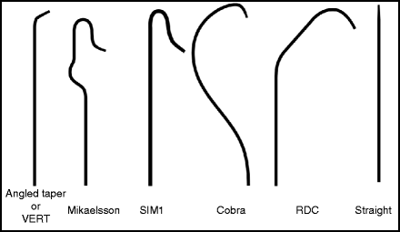
Table 3.1
Catheters for spinal angiography
Catheter | Use |
|---|---|
5F Angled Taper | Good all-purpose diagnostic catheter for supra-aortic vessels |
5F Mikaelsson | Good all-purpose catheter for intercostal and lumbar arteries |
5F Simmons 1 | Alternative to Mikaelsson |
4 or 5F Cobra | Intercostal and lumbar arteries in younger patients |
5.5F RDC | Very stable and torquable, but stiff |
5F Straight | For retrograde flush aortic runs |
Fig. 3.1
Recommended diagnostic catheters used for spinal arteriography.
3.3.7 Vessel Catheterization
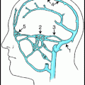
Selective spinal angiography may be either complete spinal angiography, or a partial, focused study for a specific lesion. Complete spinal angiography is a major undertaking, in which all vessels that may relate to the spinal canal are selectively catheterized and studied. This is most often used in the evaluation of a patient with a suspected dural arteriovenous fistula causing myelopathy. The vascular lesion can be anywhere from the head to the sacrum, and evaluation of all vessels supplying these structures may be required (Table 3.2). When the lesion is obviously confined to a specific region of the spine, a more focused study may be more appropriate. This should include all the vessels that supply the area of interest, and the levels above and below the lesion, given the possibility of collateral flow from adjacent spinal vessels. Another useful rule of thumb is to visualize normal spinal cord vessels above and below any lesion affecting the cord. Assessing spinal cord blood supply may require selective angiography of the vertebral arteries (Fig. 3.2), thyrocervical and costocervical trunks, subclavian arteries, intercostal arteries (Fig. 3.3), lumbar arteries (Fig. 3.4), and lateral and medial sacral arteries.
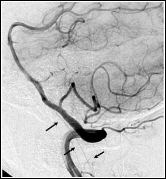
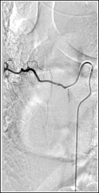
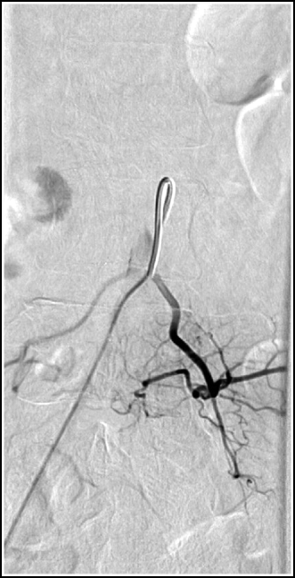
Table 3.2
Blood supply to various spinal regions
Level | Feeding arteries |
|---|---|
Upper cervical | Vertebral, ascending pharyngeal, occipital, deep cervical |
Lower cervical | Vertebral, deep cervical, ascending cervical |
Upper thoracic | Supreme intercostal, superior intercostal |
Mid-lower thoracic | Intercostal |
Upper-to-mid lumbar | Lumbar |
Lower lumbar | Iliolumbar |
Sacrum | Anterior and lateral sacral |
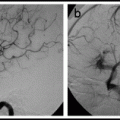
Fig. 3.2
Lateral vertebral artery angiogram showing anterior spinal artery (arrows).
Fig. 3.3
Typical intercostal artery.
Fig. 3.4
Typical lumbar artery.
3.3.8 Roadmapping
Roadmapping can be used to aid catheterization of the supra-aortic vessels, such as vertebral arteries, and the thyrocervical and costocervical trunks. Roadmapping is less helpful in catheterizing the intercostal and lumbar arteries, since respiratory motion degrades the image.
3.3.9 Double Flushing
Catheter flushing technique is discussed in Chap. 2. Although some practitioners advocate double flushing of catheters only in the supra-aortic vessels, it makes more sense to use a meticulous flushing technique anywhere in the vascular system. This ensures that one will not forget to use good technique when it is most needed. Moreover, thrombus or air emboli in spinal cord vessels can be just as disabling as cerebral ischemia.
3.3.10 Continuous Saline Infusion
Three-way stopcock or manifolds can be used to provide a heparinized saline drip through the catheter. This is particularly useful for long spinal angiographic procedures. In-line air filters (B. Braun, Bethlehem, PA) on the saline drip tubing provide added protection from bubbles (as discussed in Chap. 4). A rotating adapter on the stopcock is needed to prevent the stopcock from being a drag on free manipulation of the catheter. Using both a rotating three-way stopcock and a rotating hemostatic valve on the catheter allows for two pivot points to allow free rotation of the catheter. This is important, as the catheter may not be in a stable position in the small lumbar and intercostal arteries.
3.3.11 Hand Injection
Frequent small injections (“puffing”) of contrast can be used to help manipulate the catheter into the desired lumbar and intercostal arteries. A 20 mL syringe containing contrast can be left attached to the catheter for these injections, and then used immediately for hand injections of contrast for angiographic runs. As is done in the cerebral vasculature, the syringe is held vertically and care is taken not to allow bubbles to enter the catheter. Most spinal vessels are best imaged with hand injections of contrast, to allow for modulation of the injection rate and volume, depending on the size of the vessel and stability of the catheter. An adequate angiographic run can be usually done with a single 2–3 s injection of 4–6 mL (100% contrast) of contrast. The goal is to adequately opacify the vessel of interest without displacing the catheter or refluxing too much into the aorta or into the ever-present collaterals to other spinal vessels. Patients should be warned that they will experience warmth and/or cramping in the territory of the injected vessel, and breathing should be suspended during the angiographic run, but the phase of respiration at which the breath-holding should occur depends on the spinal level being imaged (see below).
3.3.12 Mechanical Injection
A power contrast injector is necessary for thoracic or lumbar aortic angiograms, and for large vessels such as subclavian or iliac arteries. As stated in Chap. 2, the pressure and flow rate settings should not exceed the ratings of the stopcock or catheter. Common power injector settings for vessels studied in spinal angiograms using a 5F catheter are listed in Table 3.3. Note that one may need to increase or decrease these rates and volumes, depending on the size of the vessels, the stability of the catheter, and the quickness of the runoff of the contrast on a test injection. Use extreme caution if the catheter is wedged in the vessel and be especially careful if there is a possibility that a spinal cord vessel is arising from the branch one is injecting, since high-pressure power injections can damage the cord. When in doubt, use careful hand-injections of contrast.
Table 3.3
Standard power injector settingsa
Vessel | Power injector settings |
|---|---|
Aortic arch | 20 mL/s; total of 25 mL |
Retrograde aortic flush | 10 mL/s; total of 30 mL |
Iliac artery | 10 mL/s; total of 20 mL |
Subclavian artery | 6 mL/s; total of 15 mL |
Vertebral artery | 6 mL/s; total of 8 mL |
Lumbar or intercostal artery | 2 mL/s; total of 6 mL |
For 3D imaging | 0.5–2 mL/s; total 7–30 mL (higher doses for high flow AVF) |
3.3.13 Vessel Selection
If the exact level of the lesion is known from non-invasive imaging, the spinal angiogram should begin with those vessels supplying that area. Following catheterization of the vessel of interest, it is then customary to work systematically above and below the lesion to include normal territory adjacent to the lesion. Lesions of the cord itself usually require mapping of the spinal cord supply above and below the lesion. For complete spinal angiography, it is particularly important to image the intercostal and lumbar arteries in a systematic fashion so that one does not inadvertently miss or repeat a level. It is helpful to maintain a worksheet during the procedure, and list the sides and vessels injected during each angiographic run. Radio-opaque marker rulers can be placed under the patient on the table or marker tapes can be affixed to the patient’s back, slightly off midline to have a reference available on each film to help confirm the levels studied. Additionally, bony landmarks, such as the 12th rib, can also help with keeping track of the vessels being studied.
Related posts:


Stay updated, free articles. Join our Telegram channel

Full access? Get Clinical Tree


