Cervical Spine Axial 1

Pathologic Process
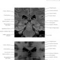
Note on these images that the nasopharynx is often seen on cervical spine imaging. The neuroradiologist should always check for symmetry of the Eustachian tube opening and fossa of Rosenmüller just anterior to the longus capitis muscle to ensure that no nasopharyngeal lesion is present (see Chapter 13 ).
Imaging Technique Consideration
Spinal imaging can be daunting at first due to the complex three-dimensional (3D) anatomy of the vertebrae and the critical implications of damage to the spine. This is particularly true taking into account various signal characteristics on magnetic resonance imaging (MRI) and motion artifact from vessel pulsation and respiration. A good principle is that computed tomography (CT) and MRI are complementary. In general, CT better delineates the architecture of the dense bones, the cortex of which does not provide much MR signal, and MRI is better at providing contrast to the soft tissues, such as the spinal cord. Although CT (top image) would be better at showing a subtle fracture of the vertebral artery foramen, MRI (bottom image) would be better at delineating hematoma in the wall of the vertebral artery from dissection.
Another good principle for learning spinal anatomy is that the lower the level in the spine, the larger the vertebrae and the easier it is to understand the anatomy. Thus it is often best to start with the lumbar spine, learning both diagnostic principles and image-guided intervention, and to work up the spine as familiarity with spinal anatomy increases.

Cervical Spine Axial 2

Diagnostic Consideration
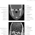
The predental space, or distance between the anterior aspect of the dens and the posterior border of the anterior arch of C1 (atlas), should measure 3 mm or less in the adult patient. Widening of this space or asymmetry of soft tissue lateral to the odontoid raises concern for cruciate ligament injury. The best imaging test is voluntary flexion and extension sequences on lateral cervical radiographs to check for change in the predental space measurement. However, in the patient who has pain limiting motion, or in the unconscious patient, CT or MRI can provide secondary signs such as fracture of C1 or C2 (axis), hemorrhage along the dura, and edema within the soft tissues.

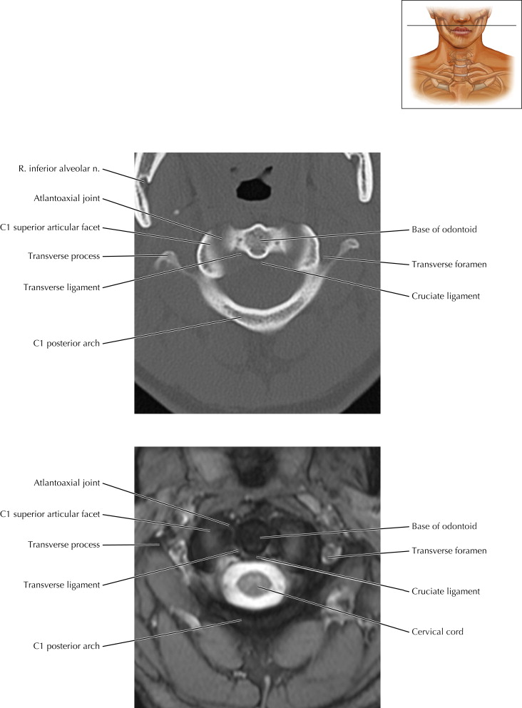
Cervical Spine Axial 3

Pathologic Process
Somewhat counterintuitively, a fracture through the larger base of the odontoid process is less serious than a fracture more superiorly near the tip. The better, more proximal blood supply at the odontoid base allows better healing and less chance of avascular necrosis.


Cervical Spine Axial 5

Imaging Technique Consideration
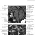
The MR images (lower) are axial 3D T2∗ gradient images. The advantages over standard 2D spin-echo sequences are the higher spatial resolution and the ability to reformat in any plane. Motion, however, tends to cause all the images of the sequence to become blurred.

Cervical Spine Axial 6

Pathologic Process
Uncovertebral osteophytes at the posterolateral aspects of the vertebral body can cause narrowing of the foramina and impingement of exiting nerve roots. Some osteophytes may be confluent with a broad-based disc-osteophyte complex across the entire posterior aspect of the vertebral body that can chronically flatten the spinal cord.

Cervical Spine Axial 7

Diagnostic Consideration
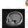
Note that the bony vertebral artery foramina, also called transverse foramina, are symmetric in this patient (Cervical Axial 7). A careful search for fracture lines across these foramina in the setting of trauma will help reveal vertebral artery dissection. Angiography (CTA or MRA) of the cervical arteries often shows some asymmetry in the lumina. If corresponding asymmetry exists in the bony foramina, the cause is likely a congenitally hypoplastic side rather than a dissection.


Cervical Spine Coronal 2

Imaging Technique Consideration
The temporomandibular joints (TMJs) can often be seen in coronal imaging of the cervical spine. In Cervical Spine Coronal 1, there is slight rotation of positioning such that the patient’s left TMJ on the right side of the image is slightly more anterior and better seen than the right TMJ.

Cervical Spine Coronal 3

Diagnostic Consideration
The coronal image is a good plane on which to assess for asymmetric positioning of the odontoid process. Note that in Cervical Spine Coronal 3 there is slightly more space to the right side of the patient’s odontoid (left side of the image), although this is likely caused by slight rotation of the patient’s neck or perhaps slight normal variation rather than trauma in this patient. The edges of the atlantoaxial joints are shown to be well aligned.

Cervical Spine Coronal 4