Ventricles and CSF Cisterns Axial 1
Normal Anatomy
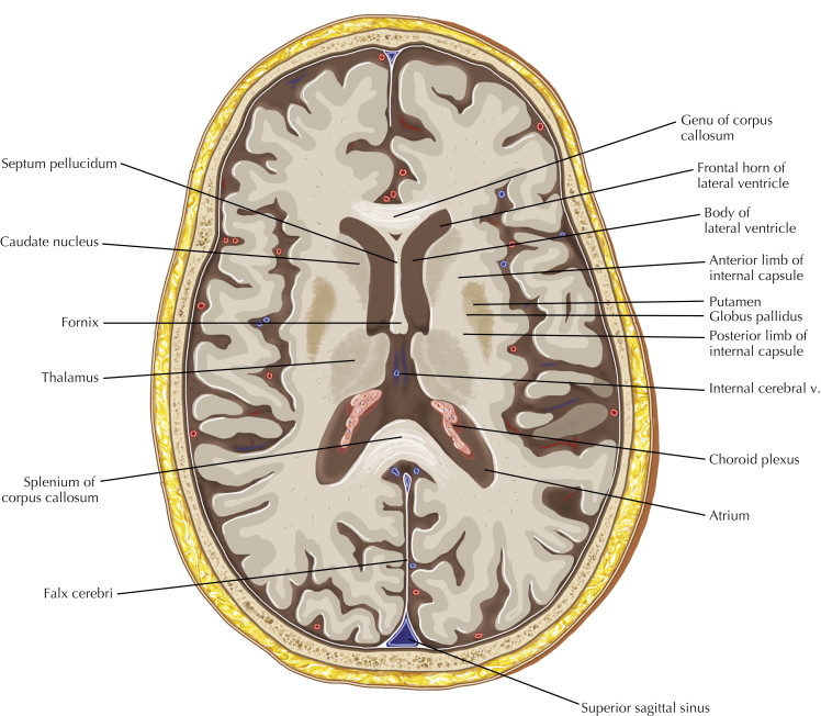
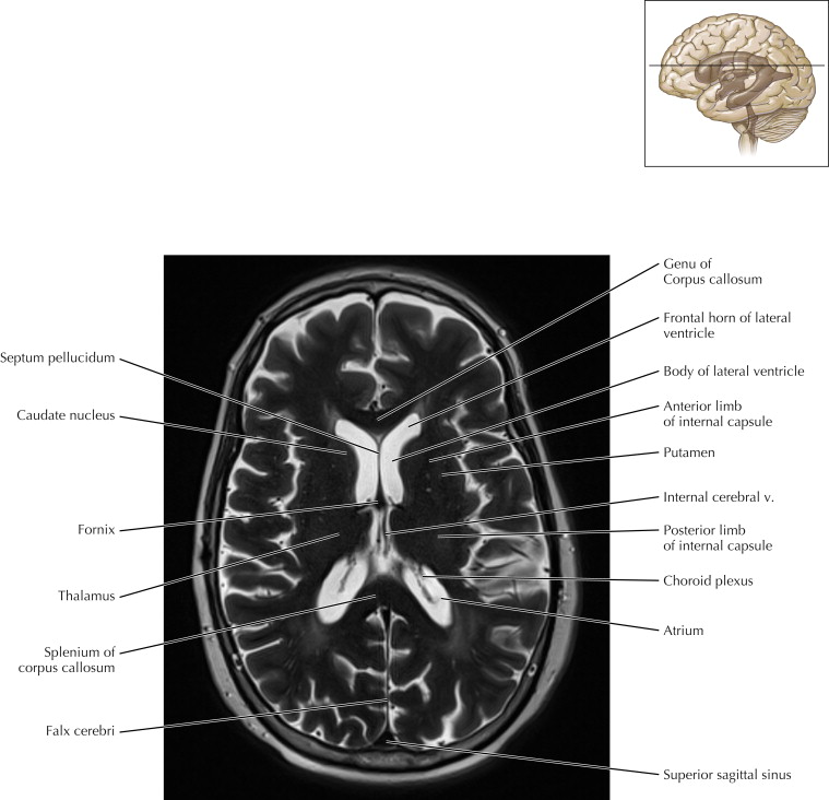
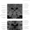
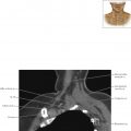
The ventricular system is located deep within the brain and is filled with cerebrospinal fluid (CSF). The normal appearance of CSF on magnetic resonance images is that of water. This axial MR image shows the choroid plexus within the ventricles, where CSF is produced. CSF is also seen outside the brain and within the sulci (subarachnoid space).
Pathologic Process
A colloid cyst is a benign lesion located near the foramen of Munro that may lead to sudden CSF obstruction, brain edema, and death if not treated with drainage and resection. CSF is produced at a rate of 500 mL daily; the intracranial vault and spinal canal contain only 150 mL, so the CSF turns over about three times each day.
Ventricles and CSF Cisterns Axial 2
Normal Anatomy
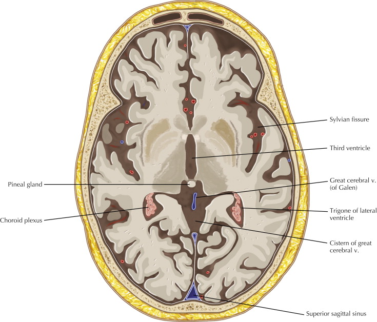
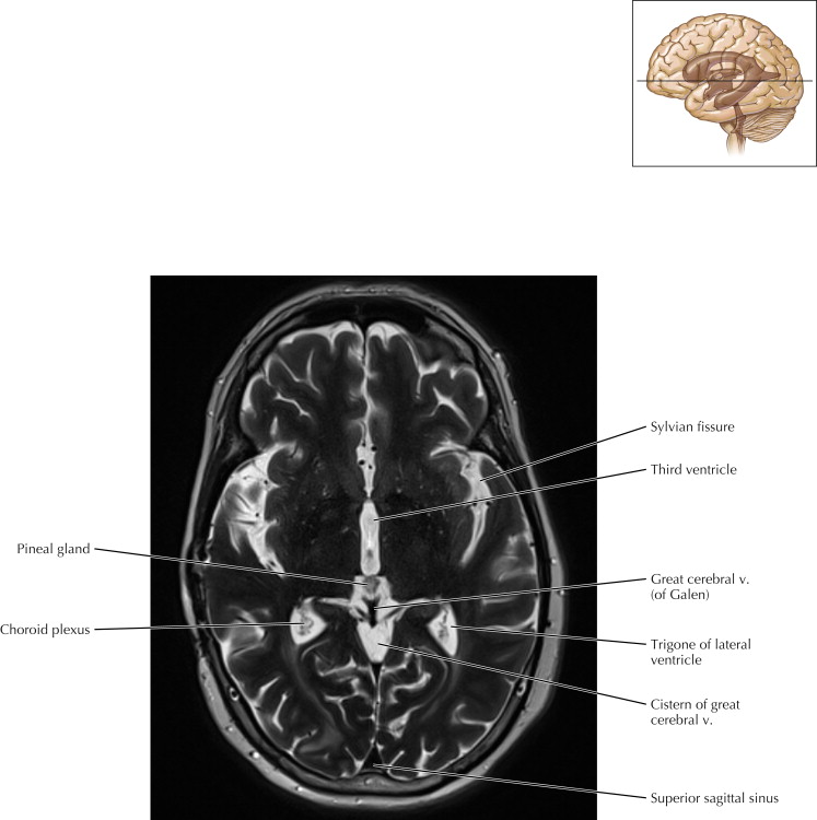
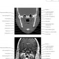
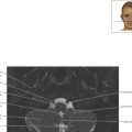
At this axial level, the atrium has a triangular appearance and thus is often referred to as the trigone.
Imaging Technique Consideration
Note that fluid is bright on T2-weighted MRI. The subcutaneous fat shows some brightness on T2-weighted images when multiple spin-echo pulses are applied (fast, or “turbo,” spin echo), as opposed to conventional spin echo T2-weighted imaging, where fat appears dark.
Ventricles and CSF Cisterns Axial 2

Normal Anatomy
At this axial level, the atrium has a triangular appearance and thus is often referred to as the trigone.
Imaging Technique Consideration
Note that fluid is bright on T2-weighted MRI. The subcutaneous fat shows some brightness on T2-weighted images when multiple spin-echo pulses are applied (fast, or “turbo,” spin echo), as opposed to conventional spin echo T2-weighted imaging, where fat appears dark.
Ventricles and CSF Cisterns Axial 3
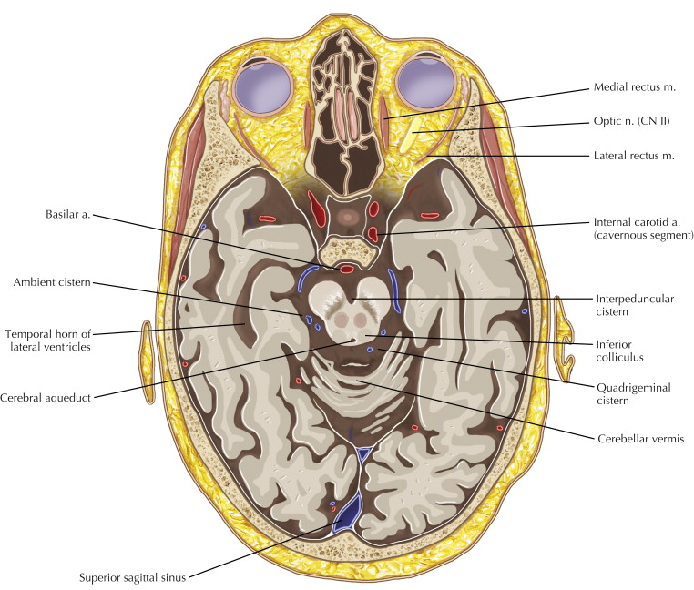
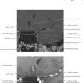
Pathologic Process
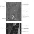
Note that the temporal horn is a contiguous part of the lateral ventricles. Dilatation of the temporal horns is frequently a sign of hydrocephalus.
Related posts:
Stay updated, free articles. Join our Telegram channel

Full access? Get Clinical Tree


